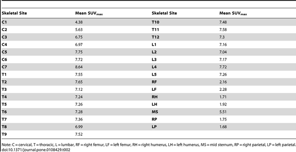
Pet scan suv values chart
At the time the article was last revised Liz Silverstone had no financial relationships to ineligible companies to disclose. The concentration of F 18 activity reflects glucose metabolism which is increased in tumor cells and inflammation.
Federal government websites often end in. The site is secure. The authors confirm that all data underlying the findings are fully available without restriction. All relevant data are within the paper. Cancer and metabolic bone diseases can alter the SUV.
Pet scan suv values chart
Cancer Imaging volume 16 , Article number: 35 Cite this article. Metrics details. Interpretation requires integration of the metabolic and anatomic findings provided by the PET and CT components which transcend the knowledge base isolated in the worlds of nuclear medicine and radiology, respectively. This encompasses how we display, threshold intensity of images and sequence our review, which are essential for accurate interpretation. For interpretation, it is important to be aware of benign variants that demonstrate high glycolytic activity, and pathologic lesions which may not be FDG-avid, and understand the physiologic and biochemical basis of these findings. This is a modality with many patterns of structural, physiologic and biochemical abnormalities that transcend the boundaries previously isolated in the worlds of nuclear medicine or radiology in characterising pathological conditions, particularly including cancer. Future articles in this series will address the use of other tracers pertinent to other cancers. Patient preparation is important in acquiring good quality studies and it is the responsibility of the PET specialist to ensure that appropriate protocols are in place to prevent non-diagnostic or suboptimal studies. Detailed discussion of acquisition parameters is beyond the scope of this review but includes preparation of diabetic patients, strategies to minimise brown fat activation, as well as prescription of the extent of the field-of-view and the positioning of the patient to address the clinical question. For example, we position the patient with their arms down for head and neck malignancies but with their arms up for thoracic cancers.
In terms of diagnosis, PET helps us pick up on abnormal behavior and figure out that something is a problem, even when it appears to be normal. Full size image. This means that more FDG is available for uptake in other tissues, including the liver.
Federal government websites often end in. The site is secure. Fusion was performed on nodes with reported having metabolic activity. The reference standard was cytology. Below SUV max of 2.
At the time the article was last revised Liz Silverstone had no financial relationships to ineligible companies to disclose. The concentration of F 18 activity reflects glucose metabolism which is increased in tumor cells and inflammation. SUV is also known as the dose uptake ratio DUR and is a mathematically derived ratio of tissue radioactivity concentration at a point in time C T at a specific region of interest ROI and the injected dose of radioactivity per kilogram of the patient's body weight 7 :. Uptake values are sometimes normalized to lean body mass LBM or body surface area. However body weight is the most commonly used because it is easy to calculate and is reproducible 7. SUV may be influenced by biological and technical factors such as blood glucose level, image noise, image resolution and variable region of interest selection. The cut off between benign and malignant lesions is in the SUV range of 2. Follow-up imaging on the same scanner and using the same method improves the reliability of serial SUV measurements. Please Note: You can also scroll through stacks with your mouse wheel or the keyboard arrow keys.
Pet scan suv values chart
The FDG is distributed throughout the body based on how much uptake there is in the tissues. Higher SUV values means there is more uptake in the tissues. This is reflected in the scan as a hotter or brighter area. The precise definition of SUV is a ratio of radioactivity concentration in tissue at a point in time divided by the injected dose of radioactivity per kilogram of the patients weight. Quite complicated but not crucial to remember for scan interpretation. SUV can change over time because of imaging factors like image noise and how the SUV is measured by the user. The SUV gives us an idea of how hot or active the tissues are compared to the rest of the body. The higher the SUV, the more likely it is abnormal.
Omegle vr
All relevant data are within the paper. J Natl Cancer Inst. Since the process of a tumor-shrinking generally takes much longer, being able to observe its behavior allows patients and doctors to find out whether or not therapy is working sooner. But accurately interpreting PET scans is complex — more so than other types of imaging. If you would like an expert second opinion on your medical imaging scan from Dr. The intensity of uptake in metastases usually parallels that in the primary site of disease. Last revised:. You can also search for this author in PubMed Google Scholar. Immediately after and without moving the patient, an emission scan was obtained in 3D mode in 11 beds at 3 minutes per bed over the same anatomical regions. It is important to review these images on a workstation that has capacity to triangulate findings in axial, coronal and sagittal planes. Eur J Nucl Med. Similarly, some aggressive sarcomas or mucinous tumours can also appear PET negative when the signal from cancer cells is dominated by the low uptake in adjacent by extra-cellular matrix or mucin production. For the L probe a bracket and an electromagnetic tracker were added. Sheikh or one of our other subspecialists, you can learn more here.
Federal government websites often end in.
For lymphoma we divide the report into nodal and extra-nodal sub-headings. This is an open-access article distributed under the terms of the Creative Commons Attribution License, which permits unrestricted use, distribution, and reproduction in any medium, provided the original author and source are properly credited. Therapeutic interventions targeting metabolic e. PET Clin. J Nucl Med 47 : — Six-month changes have to be an overall assessment. J Bone Miner Res 26 : — Results The characteristics of patients are shown in Table 1. Sheikh] Getting a second opinion allows patients to have the peace of mind that a nuclear medicine expert has reviewed their scans. With this colour scale, the liver will generally appear blue with flecks of green with adjusgment if not Fig. Figure 1. Despite the difference in SUVmax of the liver secondary to differences in weights of the two patients a and b , the liver intensity this appears the same in both patients. A separate linear tract of metabolic activity was also seen green arrow extending from the pre-sacral abnormality to the peri-anal region not shown.


0 thoughts on “Pet scan suv values chart”