Cerebellopontine angle cyst
At the time the article was last revised Ashesh Ishwarlal Ranchod had no financial relationships to ineligible companies to disclose.
Federal government websites often end in. The site is secure. Arachnoid cysts are benign developmental collections of cerebrospinal fluid CSF. The cerebellopontine angle CPA arachnoid cysts are rare and often asymptomatic. The onset of symptoms and signs is usually due to the compression of the brain, cranial nerves and obstruction of CSF circulation.
Cerebellopontine angle cyst
The cerebellopontine angle CPA is a rare location for arachnoid cysts, and fewer than 35 cases of arachnoid cysts occurring in the CPA have been reported in the literature. We discuss the diagnosis, radiographic imaging, and management of CPA arachnoid cysts. Arachnoid cysts often present with only subtle signs or symptoms, such as headache or ataxia. Our case involves a middle-aged, female patient, who presented with unilateral tinnitus, unsteadiness, and headaches associated with nausea and vomiting. On clinical examination there were no cerebellar signs or cranial neuropathy; she did, however, suffer from a unilateral mild to severe hearing loss. Recent advances in MRI magnetic resonance imaging scan techniques have led to more frequent diagnosis of CPA arachnoid cysts and with a higher degree of certainty. These lesions have a characteristic location in the posterior-inferior aspect of the CPA below the facial and vestibulocochlear nerves. Although CPA arachnoid cysts represent a small number of total arachnoid cysts, the CPA is the second most common location for arachnoid cysts to occur. The optimal surgical management of arachnoid cysts remains controversial. Most surgeons advocate that the definitive treatment for arachnoid cysts is a retrosigmoid suboccipital craniotomy and microsurgical resection and fenestration of the cyst walls. However, these cysts often do not show any change in size on repeated MRI scan and the patients' symptoms do not progress over a long period of follow-up. These clinical and radiological findings would support a conservative management approach to the majority of the arachnoid cysts. Year Archive
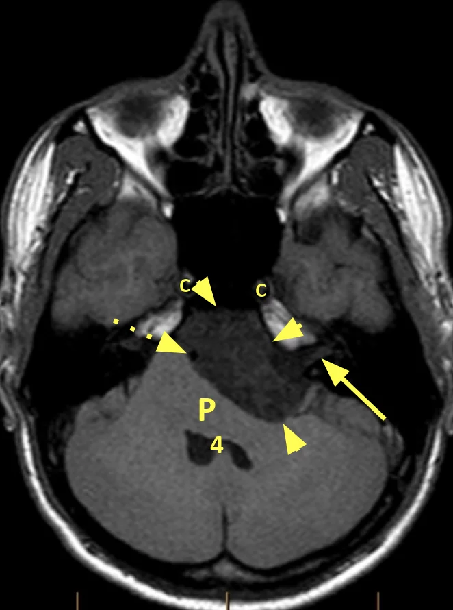
It was causing compression of right cerebellar hemisphere and brain stem.
A CSF density lesion in the right cerebellopontine angle, with moderate local mass effect with mild displacement of the brainstem but no effacement of the fourth ventricle. No widening of the adjacent porus acousticus. Remodeling of the adjacent bone is, however, present. Conclusion: Right cerebellopontine angle cystic lesion most likely represents an incidental arachnoid cyst with a differential diagnosis that includes epidermoid cyst or cystic acoustic neuroma the latter two are thought less likely. As demonstrated on the preceding CT scan, within the right cerebellopontine angle is a well-circumscribed CSF intensity lesion with bony remodeling of the petrous apex and indentation of the middle cerebellar peduncle. Content follows CSF on all sequences including diffusion-weighted imaging.
At the time the article was last revised Ashesh Ishwarlal Ranchod had no financial relationships to ineligible companies to disclose. C erebellopontine angle CPA masses are relatively common. Although a diverse range of pathologies may be seen in this region, the most common by far is vestibular schwannoma. Cerebellopontine angle masses can be divided into four groups, based on imaging characteristics:. Please Note: You can also scroll through stacks with your mouse wheel or the keyboard arrow keys. Updating… Please wait. Unable to process the form. Check for errors and try again. Thank you for updating your details. Recent Edits.
Cerebellopontine angle cyst
Federal government websites often end in. The site is secure. Arachnoid cysts are non-neoplastic, intracranial cerebrospinal fluid CSF -filled spaces lined with arachnoid membranes. Large arachnoid cysts are often symptomatic because they compress surrounding structures; therefore, they must be treated surgically. As several surgical management options exist, we explore the best approach according to each major type of arachnoid cyst: middle cranial fossa cyst, suprasellar cyst, intrahemispheric cyst, and quadrigeminal cyst. Arachnoid cysts can be classified as primary developmental cysts or secondary cysts.
100 usd to rand
At the time the case was submitted for publication Frank Gaillard had no recorded disclosures. Log in Sign up. Arachnoid cysts often present with only subtle signs or symptoms, such as headache or ataxia. Open in a separate window. Case 5: epidermoid Case 5: epidermoid. J Pediatr Neurosci. Conclusion: Although CPA arachnoid cysts represent a small number of total arachnoid cysts, the CPA is the second most common location for arachnoid cysts to occur. Close Please Note: You can also scroll through stacks with your mouse wheel or the keyboard arrow keys. By System:. The site is secure. Arachnoid cysts in the CPA are often asymptomatic. Case 3: trigeminal schwannoma Case 3: trigeminal schwannoma. Cerebellopontine angle mass.
Thank you for visiting nature. You are using a browser version with limited support for CSS.
Year Archive Remodelling of the adjacent bone is, however, present. Diagnosis certain. Conclusion: Although CPA arachnoid cysts represent a small number of total arachnoid cysts, the CPA is the second most common location for arachnoid cysts to occur. Case vestibular schwannoma Case vestibular schwannoma. Sign Up. How to use cases. Citation, DOI, disclosures and article data. Results: All patients underwent a retrosigmoid suboccipital craniotomy and microsurgical resection and fenestration of the cyst walls. Congenital mirror movements: A clue to understanding bimanual motor control. All five arachnoid cysts compressed the cerebellum or brain stem. Keywords: Arachnoid cyst, cerebellopontine angle, mirror movement. Mirror movements are involuntary movements, which occur in a muscle group or limb on one side of the body in response to an intentionally performed movement in corresponding contralateral muscle group or limb. Conflicts of interest There are no conflicts of interest. Most surgeons advocate that the definitive treatment for arachnoid cysts is a retrosigmoid suboccipital craniotomy and microsurgical resection and fenestration of the cyst walls.


It was and with me. Let's discuss this question. Here or in PM.
I to you am very obliged.