Sliding filament theory coloring answers
Sliding Filament Graphic - shows how actin and myosin interact, labeled in stages. Includes questions to answer. Sliding Filament Model slides - shows the steps of mucles contraction, students label a worksheet.
This worksheet lists the steps involved in the sliding filament model of muscle contraction and includes a coloring page of the model. Students color and answer questions. This download has two versions of the student worksheet 2 pages vs 1 pages and the answer key to the questions. Log In Join. View Wish List View Cart.
Sliding filament theory coloring answers
Each skeletal muscle is an organ that consists of various integrated tissues. These tissues include the skeletal muscle fibers, blood vessels, nerve fibers, and connective tissue. Each skeletal muscle has three layers of connective tissue called mysia that enclose it, provide structure to the muscle, and compartmentalize the muscle fibers within the muscle Figure Each muscle is wrapped in a sheath of dense, irregular connective tissue called the epimysium , which allows a muscle to contract and move powerfully while maintaining its structural integrity. The epimysium also separates muscle from other tissues and organs in the area, allowing the muscle to move independently. Inside each skeletal muscle, muscle fibers are organized into bundles, called fascicles , surrounded by a middle layer of connective tissue called the perimysium. This fascicular organization is common in muscles of the limbs; it allows the nervous system to trigger a specific movement of a muscle by activating a subset of muscle fibers within a fascicle of the muscle. Inside each fascicle, each muscle fiber is encased in a thin connective tissue layer of collagen and reticular fibers called the endomysium. The endomysium surrounds the extracellular matrix of the cells and plays a role in transferring force produced by the muscle fibers to the tendons. In skeletal muscles that work with tendons to pull on bones, the collagen in the three connective tissue layers intertwines with the collagen of a tendon. At the other end of the tendon, it fuses with the periosteum coating the bone.
Contraction of Skeletal Muscle Created by.
For complaints, use another form. Study lib. Upload document Create flashcards. Flashcards Collections. Documents Last activity. Add to
In the sliding filament model, the thick and thin filaments pass each other, shortening the sarcomere. Movement often requires the contraction of a skeletal muscle, as can be observed when the bicep muscle in the arm contracts, drawing the forearm up towards the trunk. The sliding filament model describes the process used by muscles to contract. It is a cycle of repetitive events that causes actin and myosin myofilaments to slide over each other, contracting the sarcomere and generating tension in the muscle. To understand the sliding filament model requires an understanding of sarcomere structure.
Sliding filament theory coloring answers
Sliding Filament Graphic - shows how actin and myosin interact, labeled in stages. Includes questions to answer. Sliding Filament Model slides - shows the steps of mucles contraction, students label a worksheet. Label Musscle Structure - includes entire muscle epimysium, perimysium, endomysium , then has a sarcomere labeling. Presented as Google slides, that students can drag labels to structures. Label Neuromuscular Junction - Google slide labeling that includes how a neuron interacts with a muscle motor unit. Sarcomere Coloring - black and white image shows the sarcomere and the actin and myosin filaments. Sliding Filament Model with Video - shows the motor unit and the actin and myosin interactions. Includes a video for students watch great for online learning! Includes questions to answer Sliding Filament Model slides - shows the steps of mucles contraction, students label a worksheet Label Musscle Structure - includes entire muscle epimysium, perimysium, endomysium , then has a sarcomere labeling.
Northgate property group
Algebra 2. Share this resource. I created and used this diagram while teaching Anatomy and Physiology. High school math. Character education. The myosin head will be locked in this position, attached to the actin, until another ATP molecule comes and attaches to the myosin head. Having many nuclei allows for production of the large amounts of proteins and enzymes needed for maintaining normal function of these large protein dense cells. If you've found an issue with this question, please let us know. Color the thick filaments not labeled red and the thin filaments blue. The actin filament slides inward and shortens, or contracts, the whole muscle. Also included in: Muscular System Unit Bundle. Independent work. The actin and myosin filaments slide past one another.
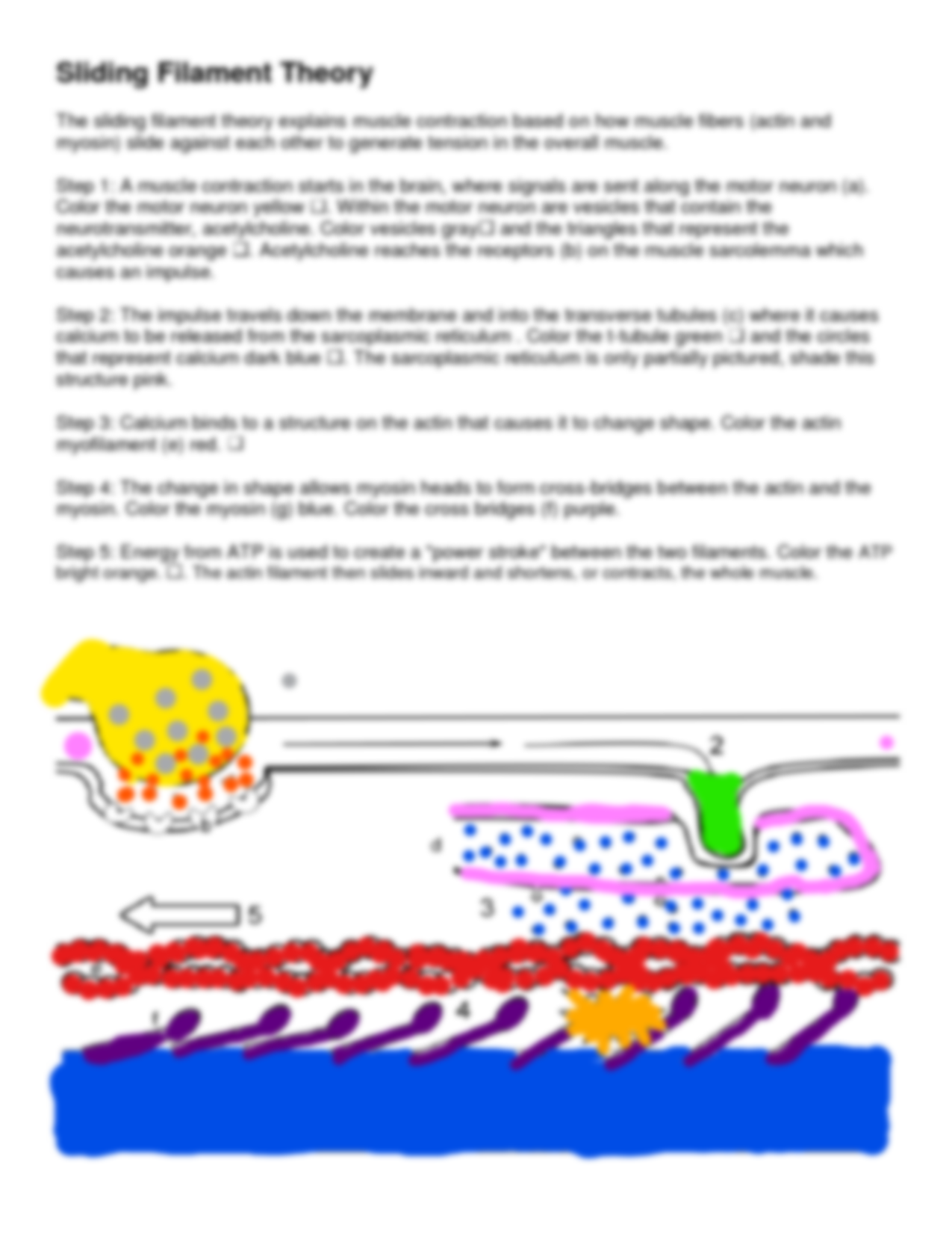
This worksheet lists the steps involved in the sliding filament model of muscle contraction and includes a coloring page of the model. Students color and answer questions.
Hispanic Heritage Month. Anatomy Muscular System quiz, sliding filament theory , muscle contraction type, muscle fibers. Just me. Color the ATP orange. We are open Saturday and Sunday! Color vesicles b gray. Physical therapy. Biology, Biology, Microbiology. Color the myosin filament m yellow. Visible to Everyone.


I think, what is it � a false way. And from it it is necessary to turn off.
Your inquiry I answer - not a problem.