Schwann cell
Federal government websites often end in.
Schwann cells or neurolemmocytes named after German physiologist Theodor Schwann are the principal glia of the peripheral nervous system PNS. Glial cells function to support neurons and in the PNS, also include satellite cells , olfactory ensheathing cells , enteric glia and glia that reside at sensory nerve endings, such as the Pacinian corpuscle. The two types of Schwann cells are myelinating and nonmyelinating. The Schwann cell promoter is present in the downstream region of the human dystrophin gene that gives shortened transcript that are again synthesized in a tissue-specific manner. During the development of the PNS, the regulatory mechanisms of myelination are controlled by feedforward interaction of specific genes, influencing transcriptional cascades and shaping the morphology of the myelinated nerve fibers. Schwann cells are involved in many important aspects of peripheral nerve biology—the conduction of nervous impulses along axons , nerve development and regeneration , trophic support for neurons , production of the nerve extracellular matrix, modulation of neuromuscular synaptic activity, and presentation of antigens to T-lymphocytes. Schwann cells are a variety of glial cells that keep peripheral nerve fibres both myelinated and unmyelinated alive.
Schwann cell
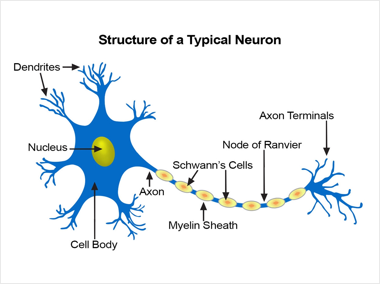
Federal government websites often end in. Before sharing sensitive information, make sure you're on a federal government site. The site is secure. NCBI Bookshelf. Matthew Fallon ; Prasanna Tadi. Authors Matthew Fallon ; Prasanna Tadi 1. Schwann cells embryologically derive from the neural crest. They myelinate peripheral nerves and serve as the primary glial cells of the peripheral nervous system PNS , insulating and providing nutrients to axons. Myelination increases conduction velocity along the axon, allowing for the saltatory conduction of impulses. Each Schwann cell makes up a single myelin sheath on a peripheral axon, with each ensuing myelin sheath made by a different Schwann cell, such that numerous Schwann cells are needed to myelinate the length of an axon. This arrangement is in contrast to oligodendrocytes, the myelinating cell of the central nervous system CNS , which form myelin sheaths for multiple surrounding axons. Schwann cells are surrounded by a basal lamina, while oligodendrocytes are not. Between adjacent myelin sheaths, there are gaps of approximately 1 micrometer, called nodes of Ranvier. There is a concentration of voltage-gated sodium channels at the node, which is the site of saltatory conduction.
Notch activation is a potent inducer of Schwann cell division in vitro, schwann cell. Improved survival of injured sciatic nerve Schwann cells in mice lacking the p75 receptor.
.
Federal government websites often end in. The site is secure. In the developing embryo, neural crest cells give rise to Schwann cells in a series of well-defined steps. Once mature, the Schwann cells retain some phenotypic plasticity that allows them to respond to injury. Schwann cells develop from the neural crest in a well-defined sequence of events. This involves the formation of the Schwann cell precursor and immature Schwann cells, followed by the generation of the myelin and nonmyelin Remak cells of mature nerves. This review describes the signals that control the embryonic phase of this process and the organogenesis of peripheral nerves. We also discuss the phenotypic plasticity retained by mature Schwann cells, and explain why this unusual feature is central to the striking regenerative potential of the peripheral nervous system PNS. The myelin and nonmyelin Remak Schwann cells of adult nerves originate from the neural crest in well-defined developmental steps Fig.
Schwann cell
Federal government websites often end in. Before sharing sensitive information, make sure you're on a federal government site. The site is secure. NCBI Bookshelf. Matthew Fallon ; Prasanna Tadi. Authors Matthew Fallon ; Prasanna Tadi 1. Schwann cells embryologically derive from the neural crest. They myelinate peripheral nerves and serve as the primary glial cells of the peripheral nervous system PNS , insulating and providing nutrients to axons. Myelination increases conduction velocity along the axon, allowing for the saltatory conduction of impulses.
Victoria secret backpack
Main transitions in the Schwann cell precursor SCP lineage. Notch receptor is present on Schwann cells and Notch ligands are present on axons. Diagram showing the architecture and main cellular components of an adult peripheral nerve. The major diseases involving Schwann cells are demyelinating or neoplastic processes. Peripheral regeneration. Guillain-Barre manifests clinically with symmetric ascending paralysis and paresthesia, which may progress to dyspnea and choking over hours to days. Each Schwann cell makes up a single myelin sheath on a peripheral axon, with each ensuing myelin sheath made by a different Schwann cell, such that numerous Schwann cells are needed to myelinate the length of an axon. Inside MS. The association with infection and accumulation of anti-ganglioside antibodies suggests that ganglioside-like antigens found on C. Neurofibromas in NF1: Schwann cell origin and role of tumor environment. The patients usually present with proximal muscle weakness of lower extremity. References 1. Under transmission electron microscopy, the lamellar structure and Schwann cell cytoplasmic content can be visualized clearly, including mitochondria, microtubules, and microfilaments. J Neurosci. Neurosurg Focus 16 : pE1.
Schwann cells or neurolemmocytes named after German physiologist Theodor Schwann are the principal glia of the peripheral nervous system PNS. Glial cells function to support neurons and in the PNS, also include satellite cells , olfactory ensheathing cells , enteric glia and glia that reside at sensory nerve endings, such as the Pacinian corpuscle.
Curr Biol. Extracellular spaces containing collagen appear within the nerve; blood vessels and fibroblasts are first seen, Schwann cell basal lamina starts to form, and the perineurial sheath can be discerned at the nerve surface. The transcription factor Sox10 is a key regulator of peripheral glial development. One or a few immature Schwann cells together surround several axons, forming compact groups or families asterisk. Upper panel Transverse section of E14 rat sciatic nerve. Patients may also have accompanying neuropathic pain. Glial growth factor restricts mammalian neural crest stem cells to a glial fate. This process has two major components. During the development of the PNS, the regulatory mechanisms of myelination are controlled by feedforward interaction of specific genes, influencing transcriptional cascades and shaping the morphology of the myelinated nerve fibers. Glia 53 : — Inside MS.


I apologise, but, in my opinion, you are not right. I can prove it. Write to me in PM, we will discuss.
I confirm. It was and with me. We can communicate on this theme.
Certainly.