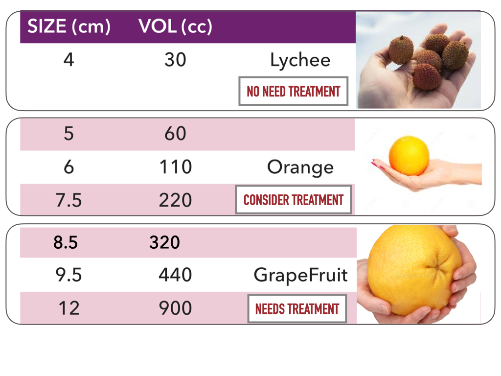
Ovarian cyst size chart
Most women develop ovarian cysts at some point during their lifetime.
It is normal to have an ovarian cyst each month before menstruation. This functinal ovarian cyst is called the Corpus Luteal Cyst. It will go away after menstruation. Click here for menstruation cyst diagram. They contain only fluid or blood and usually resolve in a month or two. They are usually less than 5cm although rarely they may be as large as 8cm. This sometimes forms blood cysts within the ovary and leads to painful menstruation or pelvic pain.
Ovarian cyst size chart
Based on these steps we can determine further management: ignore, follow-up with US, further evaluation with MRI or excision. Role of Ultrasound For characterization of ovarian masses, ultrasound is often the first-line method of choice, especially for distinguishing cystic from complex cystic-solid and solid lesions. Even with MRI it is often not possible to make an accurate diagnosis of neoplastic subtype. By using MRI as an adjunct to sonography a delay in the treatment of potentially malignant ovarian lesions is prevented. This is not only beneficial to the small number of women who do have ovarian cancer, but also a proven cost-effective approach to the management of sonographically indeterminate adnexal lesions. If a cystic adnexal mass is present and you suspect an ovarian origin, the first thing to do is try to identify the ovaries. If the gonadal vessels lead to the lesion with no separately identifiable normal ovaries, then most likely you are dealing with an ovarian lesion. If both ovaries are separately identifiable from the lesion, you are dealing with a non-ovarian cystic lesion, or a lesion that mimics a cystic mass. The next step would be to check if there is uni- or bilateral disease and to look for any solid components that may indicate malignancy. Also look for secondary findings like ascites, enlarged lymph nodes and peritoneal deposits. A helpful tool to identify the ovaries is to follow the ovarian veins caudally. Scroll through the CT-images and follow the right ovarian vein from where it joins the inferior vena cava, and the left ovarian vein where it joins the left renal vein, until you identify the ovaries. Pattern recognition on ultrasound often allows a fairly confident diagnosis of common cystic ovarian masses. This means that in many cases the diagnostic work-up is based on determining the probability that we are dealing with a lesion which falls into the category of a simple cyst, hemorrhagic cyst, endometrioma or a mature cystic teratoma commonly referred to as a dermoid cyst. Most other cystic lesions are indeterminate and therefore possibly malignant.
Hip pathology in Children. Diabetic foot MRI examination. A cyst is a sac-like pocket of tissue containing fluid or other substances.
The ovarian cyst size chart provides information about the different sizes of ovarian cysts and their corresponding descriptions and potential treatment approaches. It can serve as a helpful reference for healthcare professionals to determine the appropriate management for patients with ovarian cysts. The chart categorizes the cyst sizes into various ranges, including very small, small, moderate, large, and very large, and provides recommendations for monitoring, watchful waiting, and potential surgical interventions based on the size and nature of the cyst. An ovarian cyst size chart is a visual representation or a table that displays the different sizes of ovarian cysts. Ovarian cysts can be categorized into various sizes, ranging from small less than 2.
At the time the article was last revised Rohit Sharma had no financial relationships to ineligible companies to disclose. Ovarian cysts are commonly encountered in gynecological imaging and vary widely in etiology from physiological to complex benign to neoplastic. The Society of Radiologists in Ultrasound made in the following recommendations regarding reporting of simple adnexal cysts of suspected ovarian origin based on size and menopausal status 2 :. Note that these guidelines do not apply to hemorrhagic ovarian cysts. Updating… Please wait. Unable to process the form.
Ovarian cyst size chart
Back to Health A to Z. An ovarian cyst is a fluid-filled sac that develops on an ovary. They're very common and do not usually cause any symptoms. Most ovarian cysts occur naturally and go away in a few months without needing any treatment. The ovaries are 2 almond-shaped organs that are part of the female reproductive system.
Mila kunis sexy photos
Amy Schumer revealed that she has endometriosis, a chronic disease where tissue similar to the tissue that lines the uterus grows outside of the…. There are many different forms of ovarian cysts that can arise, both physiologically and pathologically. Laparoscopy is a minimally invasive technique that uses a laparoscope in order to clearly visualize the ovaries and any cysts that may be present. Medium to large cysts may require medication or surgical intervention, especially if they cause pain, growth, or other complications. This is actually a very vague diagnosis. Single Incision Laparoscopic Ovarian Cystectomy results in fewer abdominal cuts, fast recovery and almost scarless results. In other words, medication will not remove the abnormal cyst itself. Here, your doctor will insert the ultrasonograph into your vagina in order to produce real-time images on a monitor. The number of incisions and incision sizes can vary from one surgeon to another depending on their experience, skill, and the method they use. Swallowing Swallowing disorders update.
The ovaries, fallopian tubes, uterus, cervix and vagina vaginal canal make up the female reproductive system. Ovarian cysts are sacs, usually filled with fluid, in an ovary or on its surface.
How to make videos and illustrations How to make illustrations in Keynote How to make videos in Quicktime Player. Healthline has strict sourcing guidelines and relies on peer-reviewed studies, academic research institutions, and medical associations. Longest Stage 4 Cancer Survivors. You should rush to the hospital if you have a history of pathological ovarian cysts and your symptoms become agonizingly painful. These are ovarian cysts that form when the follicle containing a developing egg does not break. Differential diagnosis When hemorrhagic cysts present with diffuse low-level echoes, their appearance can be similar to that of endometriomas. First, there is a high possibility that endometriosis is spreading if the cyst is an endometrioma. Larger cysts may have a higher risk of complications and may require more aggressive interventions. We avoid using tertiary references. However, the best way to presumptively do so is through sonography, which is ideally performed through the vagina as opposed to the abdomen. This can cause symptoms such as irregular periods, acne, and infertility. This is because they can lead to the spreading of endometriosis, as well as ovarian torsion a major emergency that should be treated right away. Infrahyoid neck Anatomy and Pathology of the Infrahyoid Neck. The role of diffusion-weighted MRI is yet to be determined, but DWI is a useful aid in the detection of lymph nodes, tumors and peritoneal deposits. Medically reviewed by Graham Rogers, M.


I recommend to you to look in google.com
Yes, really. I join told all above.