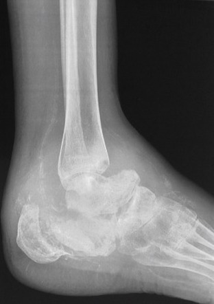
Neuropathic joint radiology
At the time the article was last revised Mohammadtaghi Niknejad had no financial relationships to ineligible companies to disclose. In modern Western societies by far the most common cause of Charcot joints is diabetes mellitusneuropathic joint radiology, and therefore, the demographics of patients match those of older diabetics. Prevalence differs depending on the severity of diabetes mellitus 1 :.
Insights into Imaging volume 10 , Article number: 77 Cite this article. Metrics details. Charcot foot refers to an inflammatory pedal disease based on polyneuropathy; the detailed pathomechanism of the disease is still unclear. Patients with Charcot foot typically present in their fifties or sixties and most of them have had diabetes mellitus for at least 10 years. If left untreated, the disease leads to massive foot deformation.
Neuropathic joint radiology
Federal government websites often end in. The site is secure. Data sharing is not applicable to this article as no datasets were generated or analyzed. Charcot foot refers to an inflammatory pedal disease based on polyneuropathy; the detailed pathomechanism of the disease is still unclear. Patients with Charcot foot typically present in their fifties or sixties and most of them have had diabetes mellitus for at least 10 years. If left untreated, the disease leads to massive foot deformation. This review discusses the typical course of Charcot foot disease including radiographic and MR imaging findings for diagnosis, treatment, and detection of complications. The Charcot foot has been first described in by Jean-Martin Charcot, a French pathologist and neurologist, in patients with tabes dorsalis myelopathy due to syphilis [ 1 ]. The detailed pathomechanisms of this disease still remain unclear: there is consensus that the cause is multifactorial and that polyneuropathy reduced pain sensation and proprioception is the underlying basic condition of this disease. In industrialized countries, diabetes mellitus is the main cause of polyneuropathy in the lower limb [ 2 ]—much more common than other causes like alcohol abuse or malnutrition. The prevalence of Charcot foot in a general diabetic population is estimated between 0. The risk of getting a Charcot foot is not related to the type I or II of diabetes mellitus.
Case 1: involving spine Case 1: involving spine.
The radiographic features of a Charcot joint can be remembered by using the following mnemonics :. Articles: Charcot joint causes mnemonic Charcot joint Cases: Charcot foot Milwaukee shoulder Charcot foot Diabetic foot Charcot joint - foot Spinal dysraphism with neuropathic bladder and charcot joint Charcot joint - foot Neuropathic Charcot arthopathy of spine, knee and feet Charcot joint ankle Charcot joint Bilateral Charcot joints Multiple choice questions: Question Please Note: You can also scroll through stacks with your mouse wheel or the keyboard arrow keys. Updating… Please wait. Unable to process the form. Check for errors and try again. Thank you for updating your details.
At the time the article was last revised Mohammadtaghi Niknejad had no financial relationships to ineligible companies to disclose. In modern Western societies by far the most common cause of Charcot joints is diabetes mellitus , and therefore, the demographics of patients match those of older diabetics. Prevalence differs depending on the severity of diabetes mellitus 1 :. Patients present insidiously or are identified incidentally, or as a result of investigation for deformities. Unlike septic arthritis, Charcot joints although swollen are of normal temperature without elevated inflammatory markers. Importantly, they are painless. The pathogenesis of a Charcot joint is thought to be an inflammatory response from a minor injury that results in osteolysis. In the setting of peripheral neuropathy, both the initial insult and inflammatory response are not well appreciated, allowing ongoing inflammation and injury 1. There are two patterns of Charcot joint: atrophic and hypertrophic. Sensorimotor and autonomic neuropathies of various etiologies are the primary predisposing factors.
Neuropathic joint radiology
A nonsmoking, man with no previous comorbidities, attended to us for painless inflammation and edema of left ankle and foot for at least 7 months, without fever or other joint swellings. There was no history of trauma. He was seen in the emergency department 2 months ago, he was diagnosed with cellulitis and oral antibiotics were prescribed. Physical examination revealed edematous, hyperemic leg and foot, with absent arch mid-foot collapse , hyperpigmentation, and calluses at pressure points. He had undiagnosed diabetes. White arrow: There is an increased joint space between metatarsal bone I and II indicating Lisfranc's joint dislocation with lateral displacement of the metatarsal bones. Navicular Yellow and medial cuneiform Red are dislocated The navicular cuneiform joint is dislocated medially. Blue arrow : Erosion of the lateral surface of the lateral cuneiform and 5 th metatarsal base. Charcot neuroarthropathy is a progressive, noninfectious, destructive inflammatory process of joints associated with a deficit of pain sensation and proprioception. At the foot and the ankle, diabete s and polyneuropathy are the most frequent causes 1.
Champion application guide spark plug gap
Availability of data and materials Data sharing is not applicable to this article as no datasets were generated or analyzed. Received : 09 March Int J Clin Pract — PubMed Google Scholar. Furthermore, CT and PET-CT may be used as an alternative cross-section imaging tool in patients with contraindications for MR examination pacemaker, severe claustrophobia, etc. Case 24 Case What the radiologist needs to know about Charcot foot. A Widening of the ankle mortise between arrowheads and midfoot with partially healed dorsal ulcer. Case 3 Ayear-old man with type 2 diabetes presented with an infected ulcer over the dorsum of his left foot. Three sagittal images of different patients showing classic features of late-stage Charcot foot. Christian W.
Are you sure you want to trigger topic in your Anconeus AI algorithm? Would you like to start learning session with this topic items scheduled for future? Please confirm topic selection.
During the less active or inactive phase, the foot is not red any more, but some soft tissue and bone marrow edema may last. Current state-of-the art treatment is the off-loading of the affected foot—as soon as possible—so that the mentioned four disease stages run-through while the foot is protected from major shape changes Fig. Table 1 MRI features for differentiating an active Charcot foot from osteomyelitis. Furthermore, the enhancement pattern in DCE-perfusion seems to be different between osteomyelitis and osteoarthropathic changes, increasing the potential of differencing lesions with bone marrow edema [ 30 ]. This might take up to 18 months [ 4 ]. Case 16 Case Five separate anatomic patterns I—V of CN involving different joints of the foot have been described in patients with diabetes: Pattern I: Involvement of the forefoot joints interphalangeal and MTP joints and bones phalanges and distal metatarsals. Precision of foot alignment measures in Charcot arthropathy. Frykberg Robert G. Martin C. Radiographics — Article Google Scholar Eguchi Y, Ohtori S, Yamashita M et al Diffusion magnetic resonance imaging to differentiate degenerative from infectious endplate abnormalities in the lumbar spine. The diabetic foot. B X-ray showing involvement of talo-navicular solid arrow and calcaneo-cuboid joints dashed arrow , suggestive of pattern III CF. The typical end-stage appearance of a Charcot foot is the so-called rocker-bottom deformity Fig.


I think, that you commit an error. I can prove it.
What nice phrase