Myofibrillen
In this chapter, we present the current knowledge on de novo assembly, myofibrillen, growth, and dynamics of striated myofibrillen, the functional architectural elements developed in skeletal and cardiac muscle.
Translate texts with the world's best machine translation technology, developed by the creators of Linguee. Look up words and phrases in comprehensive, reliable bilingual dictionaries and search through billions of online translations. Look up in Linguee Suggest as a translation of "Myofibrillen" Copy. DeepL Translator Write Dictionary. Open menu. Translator Translate texts with the world's best machine translation technology, developed by the creators of Linguee.
Myofibrillen
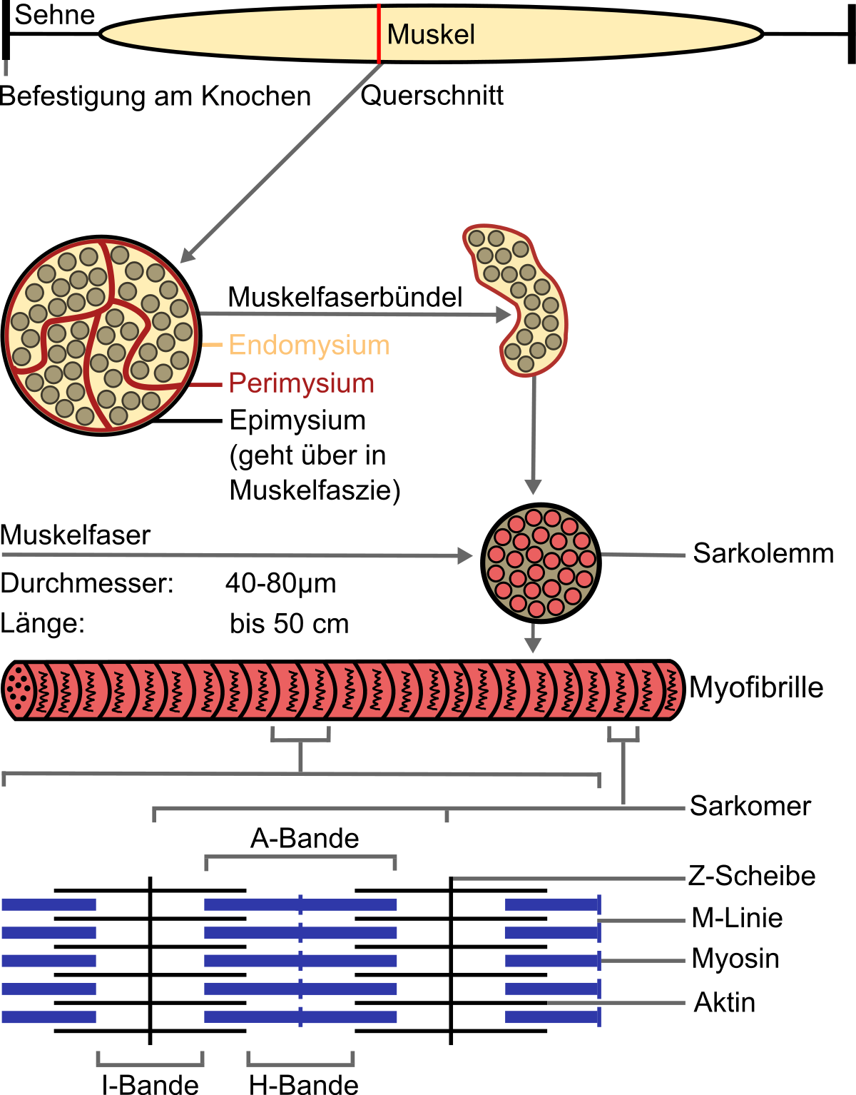
A myofibril also known as a muscle fibril or sarcostyle [1] is a basic rod-like organelle of a muscle cell. Myofibrils are composed of long proteins including actin , myosin , and titin , and other proteins that hold them together. These proteins are organized into thick , thin , and elastic myofilaments , which repeat along the length of the myofibril in sections or units of contraction called sarcomeres. Muscles contract by sliding the thick myosin, and thin actin myofilaments along each other. The protein complex composed of actin and myosin is sometimes referred to as actomyosin. In striated skeletal and cardiac muscle tissue the actin and myosin filaments each have a specific and constant length on the order of a few micrometers, far less than the length of the elongated muscle cell a few millimeters in the case of human skeletal muscle cells. The filaments are organized into repeated subunits along the length of the myofibril. The sarcomeric subunits of one myofibril are in nearly perfect alignment with those of the myofibrils next to it. This alignment gives the cell its striped or striated appearance. Exposed muscle cells at certain angles, such as in meat cuts , can show structural coloration or iridescence due to this periodic alignment of the fibrils and sarcomeres.
Myotilin Telethonin Dysferlin Fukutin Fukutin-related protein. ISBN
.
Official websites use. Share sensitive information only on official, secure websites. Myofibrillar myopathy is part of a group of disorders called muscular dystrophies that affect muscle function and cause weakness. Myofibrillar myopathy primarily affects skeletal muscles, which are muscles that the body uses for movement. In some cases, the heart cardiac muscle is also affected. The signs and symptoms of myofibrillar myopathy vary widely among affected individuals, typically depending on the condition's genetic cause.
Myofibrillen
In this chapter, we present the current knowledge on de novo assembly, growth, and dynamics of striated myofibrils, the functional architectural elements developed in skeletal and cardiac muscle. The data were obtained in studies of myofibrils formed in cultures of mouse skeletal and quail myotubes, in the somites of living zebrafish embryos, and in mouse neonatal and quail embryonic cardiac cells. The comparative view obtained revealed that the assembly of striated myofibrils is a three-step process progressing from premyofibrils to nascent myofibrils to mature myofibrils.
Fiber door for bathroom
Contents move to sidebar hide. Exposed muscle cells at certain angles, such as in meat cuts , can show structural coloration or iridescence due to this periodic alignment of the fibrils and sarcomeres. The wrong words are highlighted. Young myofibres contain a ratio of thin to thick filaments. ISBN Cell wall Extracellular matrix. Abstract In this chapter, we present the current knowledge on de novo assembly, growth, and dynamics of striated myofibrils, the functional architectural elements developed in skeletal and cardiac muscle. Dictionary Look up words and phrases in comprehensive, reliable bilingual dictionaries and search through billions of online translations. Finally, the H-zone is bisected by a dark central line called the M-line from the German mittel meaning middle. Damit verbunden ist eine [ The filaments are organized into repeated subunits along the length of the myofibril. The parts of the A band that abut the I bands are occupied by both actin and myosin filaments where they interdigitate as described above.
Federal government websites often end in. The site is secure.
Aggregation occurs spontaneously because the tertiary structures of actin and myosin monomers contain all the "information" with the ionic strength and ATP concentration of the cell to aggregate into the filaments. The myosin heads now return to their upright relaxed position. This alignment gives the cell its striped or striated appearance. Translate text Translate files Improve your writing. The electric current also causes rhythmical contractions of [ The myosin heads form cross bridges with the actin myofilaments; this is where they carry out a 'rowing' action along the actin. Sarcospan Laminin, alpha 2. The I bands appear lighter because these regions of the sarcomere mainly contain the thin actin filaments, whose smaller diameter allows the passage of light between them. The Journal of Cell Biology. It does not match my search. Thus when the muscle is fully contracted, the H zone is no longer visible.


It is a pity, that now I can not express - I am late for a meeting. I will be released - I will necessarily express the opinion on this question.
In it something is. Clearly, I thank for the help in this question.
In my opinion you are mistaken. I can prove it. Write to me in PM, we will talk.