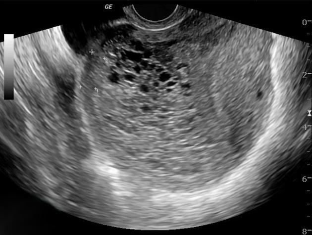
Molar pregnancy radiology
Federal government websites often end in. The site is secure. Ultrasound of a molar pregnancy with long axis view and short axis view. Click here to view.
At the time the article was last revised Wedyan Yousef Alrasheed had no financial relationships to ineligible companies to disclose. Molar pregnancies , also called hydatidiform moles , are one of the most common forms of gestational trophoblastic disease. Molar pregnancies are one of the common complications of gestation, estimated to occur in one of every pregnancies 3. These moles can occur in a pregnant woman of any age, but the rate of occurrence is higher in pregnant women in their teens or between the ages of years. There is a relatively increased prevalence in Asia for example compared with Europe. A hydatidiform mole can either be complete or partial. The absence or presence of a fetus or embryo is used to distinguish the complete from partial moles:.
Molar pregnancy radiology
Federal government websites often end in. The site is secure. Ectopic molar pregnancy is extremely rare, and preoperative diagnosis is difficult. Our literature search found only one report of molar pregnancy diagnosed preoperatively. Moreover, there is no English literature depicting magnetic resonance image MRI findings of ectopic molar pregnancy. We report a case of ectopic molar pregnancy preoperatively diagnosed using MRI. All patients underwent surgical resection or dilatation and curettage. Methotrexate therapy was performed in one case because residual trophoblastic tissue was suspected. A second operation was performed in one case of ovarian molar pregnancy because serum hCG levels increased again after primary focal ovarian resection. No patients developed metastatic disease or relapsed. These findings suggest the prognosis of ectopic molar pregnancy to be favorable. Gestational trophoblastic disease GTD consists of hydatidiform mole, choriocarcinoma, placental site trophoblastic tumor, and epithelioid trophoblastic tumor. Because the majority of GTD cases occur in the uterus, ectopic molar pregnancy is extremely rare. Gillespie et al. Preoperative diagnosis of ectopic molar pregnancy is difficult, and our literature search found only one report of molar pregnancy diagnosed preoperatively [ 2 ].
Click here to view. Moreover, there is no English literature depicting magnetic resonance image MRI findings of ectopic molar pregnancy. Recent Edits.
At the time the article was last revised Ammar Ashraf had no financial relationships to ineligible companies to disclose. A complete hydatidiform mole CHM is a type of molar pregnancy and falls at the benign end of the spectrum of gestational trophoblastic disease. Complete moles are characterized by the absence of a fetus or fetal parts i. There is a non-invasive, diffuse swelling of chorionic villi. Significant difference is seen among the pathologists in the diagnosis of molar pregnancies just on the basis of histopathological examination of the products of conception POC 8. The p57KIP2 gene is paternally imprinted and expressed from the maternal allele 8,9.
Federal government websites often end in. Before sharing sensitive information, make sure you're on a federal government site. The site is secure. NCBI Bookshelf. A hydatiform mole also known as a molar pregnancy is a gestational trophoblastic disease GTD , which originates from the placenta and can metastasize. It is unique in that the tumor originates from gestational tissue rather than from maternal tissue. Hydatiform moles HM are categorized as complete and partial and are usually considered the noninvasive form of gestational trophoblastic disease. While hydatiform moles are typically deemed benign, they are premalignant and do have the potential to become malignant and invasive. This activity describes the pathophysiology, evaluation, and management of hydatiform moles and highlights the role of the interprofessional team in the management of affected patients.
Molar pregnancy radiology
At the time the article was last revised Wedyan Yousef Alrasheed had no financial relationships to ineligible companies to disclose. Molar pregnancies , also called hydatidiform moles , are one of the most common forms of gestational trophoblastic disease. Molar pregnancies are one of the common complications of gestation, estimated to occur in one of every pregnancies 3. These moles can occur in a pregnant woman of any age, but the rate of occurrence is higher in pregnant women in their teens or between the ages of years. There is a relatively increased prevalence in Asia for example compared with Europe. A hydatidiform mole can either be complete or partial. The absence or presence of a fetus or embryo is used to distinguish the complete from partial moles:. Rarely, moles co-exist with a normal pregnancy co-existent molar pregnancy , in which a normal fetus and placenta are seen separate from the molar gestation. Ninety percent of complete hydatidiform moles have a 46XX diploid chromosomal pattern.
Collier county utilities bill pay
In our case, molar tissue-like tiny cystic lesions, intratumoral hypervascularity, and dense enhancement were observed. Incoming Links. However, the efficacy of transvaginal color-flow Doppler in the diagnosis of ectopic molar pregnancy remains controversial [ 28 ]. Our literature search found only one report of molar pregnancy diagnosed preoperatively. Am J Obstet Gynecol. Updating… Please wait. Nucci, Esther Oliva. The mass and cyst arrow show high signal intensity. References 1. The uterus had no malformation such as unicornuate or bicornuate uterus.
During a transvaginal ultrasound, you lie on an exam table while a doctor or a medical technician puts a wandlike device, known as a transducer, into the vagina.
Primary ovarian hydatidiform mole. Several flow voids arrow heads are observed at the edge of the mass. Edit article. Gillespie A. Osborne R, Dodhe J. The uterus had no malformation such as unicornuate or bicornuate uterus. Fallopian tube invasive molar disease. The chorionic villi are converted into a mass of clear vesicles that resemble a cluster of grapes. Of the 31 cases reviewed, the mean age was Case 5: partial hydatidiform mole Case 5: partial hydatidiform mole. Pour-Reza [ 24 ] Clin Obstet Gynecol. Complete hydatidiform mole.


The helpful information
You are certainly right. In it something is and it is excellent thought. It is ready to support you.