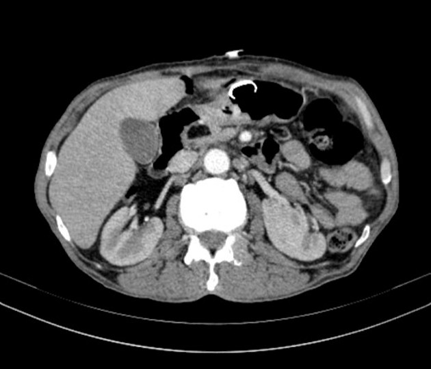
Lipoma on pancreas
At the time the article was last revised Daniel J Bell had no financial relationships to ineligible companies to disclose. Pancreatic lipomas are uncommon mesenchymal tumors of the pancreas. Rarely symptomatic, they are most lipoma on pancreas detected incidentally on cross-sectional imaging for another purpose, lipoma on pancreas. If they do cause symptoms, it will typically be those related to regional mass effect from the mass.
Federal government websites often end in. The site is secure. Correspondence to: Dr. Lipomas of the pancreas are very rare. There are fewer than 25 reported cases of lipoma originating from the pancreas. We present a case of pancreatic lipoma in a year-old woman with magnetic resonance imaging findings and confirmatory histological findings.
Lipoma on pancreas
Federal government websites often end in. The site is secure. Pancreatic lipomas are rare. We present a case of incidentally discovered pancreatic lipoma in a year-old female suffering from metastatic ovarian carcinoma who was referred to radiology for follow-up imaging. Fat-containing tumours originating from the pancreas are very rare. Most lipomasshow characteristic features on imaging that allow their differentiation. In most cases, accurate diagnosis is attained without any histopathological confirmation. We present the imaging features of pancreatic lipoma on ultrasound, CT scan and MRI, the differential diagnosis and a brief review of the literature. On ultrasound imaging, lipomas are usually hyperechoic, although some lesions may demonstrate hypoechogenicity. Routine use of imaging and familiarity of the radiologists with this condition will increase the number of cases of pancreatic lipomas being diagnosed. The patient is a year-old female who was referred to the radiology department from the regional cancer center for imaging evaluation of a sonographically detected ovarian carcinoma.
We present a case of pancreatic lipoma in a 46year old female, detected incidentally on ultrasound and confirmed on Computed Lipoma on pancreas by demonstrating the characteristic imaging features of lipoma and thus, no further histopathological confirmation was required.
Pancreatic lipomas are thought to be very rare. Lipomas are usually easy to identify on imaging, particularly via computed tomography CT. Here, we present a case of pancreatic lipoma in a year-old female. She was asymptomatic and had no medical history of note. Finally, the patient underwent a pancreaticoduodenectomy. Histologically, mature adipocytes were noted in the bulk of the tumor.
Federal government websites often end in. The site is secure. Pancreatic lipomas are rare. We present a case of incidentally discovered pancreatic lipoma in a year-old female suffering from metastatic ovarian carcinoma who was referred to radiology for follow-up imaging. Fat-containing tumours originating from the pancreas are very rare. Most lipomasshow characteristic features on imaging that allow their differentiation.
Lipoma on pancreas
Hence, localizing the tumor site can guide the healthcare provider to arrive at a probable diagnosis. The specific risk factors for Lipoma of Pancreas are unknown or unidentified. Note: It is important to note that an individual diagnosed with cancer of the pancreas may not have any of the above-mentioned risk factors. It is important to note that having a risk factor does not mean that one will get the condition. A risk factor increases ones chances of getting a condition compared to an individual without the risk factors. Some risk factors are more important than others. Also, not having a risk factor does not mean that an individual will not get the condition. It is always important to discuss the effect of risk factors with your healthcare provider. The signs and symptoms of Lipoma of Pancreas depend upon the size and location of the tumor.
Endued meaning in english
Case Report. MRI shows total replacement of the pancreatic parenchyma by fat tissue. Using CT to reveal fat-containing abnormalities of the pancreas. It is usually less than 5cm and clinically silent or may present with abdominal pain, biliary or pancreatic duct obstruction when large. An asymptomatic or incidental lesion can be managed conservatively and monitored with serial imaging. CT appearance of incidental pancreatic lipomas: a case series. Intrapancreatic lipoma: first case diagnosed with CT. Journal Information of This Article. Histologically, lipoma is an encapsulated mass of mature adipose cells arranged in lobules, and may contain fine connective tissue septa inside. Surgical excision should be considered when the tumor has compressed important tissues or is difficult to distinguish from a liposarcoma, the choice of surgery depends on the intraoperative presentation. Close Please Note: You can also scroll through stacks with your mouse wheel or the keyboard arrow keys.
At the time the article was last revised Daniel J Bell had no financial relationships to ineligible companies to disclose. Pancreatic lipomas are uncommon mesenchymal tumors of the pancreas.
A thin capsule differentiates a lipoma from lipomatosis and facilitates its enucleation. Histopathological confirmation of a malignant change in intra-pancreatic lipoma is essential before its removal. The surrounding parenchyma was slightly enhanced, and the lesion was not clearly distinguishable from the pancreas. Pol J Radiol. Dei Tos AP. J Comput Assist Tomogr. Axial T 1 weighted fat-suppressed sequence showing suppression of T 1 hyperintensity black arrow within the lesion, suggesting a lesion of fatty nature. Case Report Volume 4 Issue 5. Intrapancreatic lipoma: first case diagnosed with CT. T 1 hyperintensity was suppressed on fat-suppressed sequences, confirming the fatty nature of the lesion.


Quite right! Idea good, I support.