Hilum nedir tıp
Background: Although several studies were performed for the classification of renal venous system, abnormal venous drainage pathways were so few mentioned in literature.
Vikipediya, azad ensiklopediya. PLoS Med. Aggregation of cancer among relatives of never-smoking lung cancer patients. Int J Cancer. The accumulated evidence on lung cancer and environmental tobacco smoke.
Hilum nedir tıp
Sol 5. A year-old male patient was admitted to our clinic with complaints of shortness of breath and fatigue for 1 month. It was learned in his professional history that he had been a dental technician for 30 years. On thoracic computed tomography CT , enlarged lymph nodes and lymphadenopathies LAP with local conglomeration and calcifications in the mediastinal, subcarinal and bilateral hilar areas were seen. Widely disseminated inhomogeneous mass-like consolidation areas, including internal calcifications, extending from the hilum to the parenchyma in both lungs, were observed more prominently in the right middle zone. Diffuse interstitial thickenings, infiltrations and centrilobular nodular density increases in both lungs, nodular consolidated areas with recessed contours and nodules and ground glass densities were observed, especially in the left upper zone. In the lateral part of the left 5th rib, a heterogeneous soft tissue mass of approximately 12x5 cm, causing cortical destruction, invading the surrounding soft tissues and muscle planes, and containing internal cystic-necrotic components was observed. On abdominal CT, enlarged lymph nodes and LAPs, some of which contain calcifications, were observed in the abdomen. The pathology result was reported as Diffuse B-Cell Lymphoma in the patient who underwent transthoracic biopsy due to radiographic appearances that are not typical for PMF. Here, we presented a case of pneumoconiosis with occupational carcinogen exposure and presenting with lymphoma. Olgu Sunumu. TR EN. LaDou, J.
Philadelphia: Churchill Livingstone Elsevier. The neck of pancreas lies on the transpyloric plane, whilst the body and tail are to the left and above it.
The transpyloric plane , also known as Addison's plane , is an imaginary horizontal plane , located halfway between the suprasternal notch of the manubrium and the upper border of the symphysis pubis at the level of the first lumbar vertebrae, L1. It lies roughly a hand's breadth beneath the xiphisternum [1] or midway between the xiphisternum and the umbilicus. The transpyloric plane is clinically notable because it passes through several important abdominal structures. It also divides the supracolic and infracolic compartments , with the liver, spleen and gastric fundus above it and the small intestine and colon below it. The first lumbar vertebra lies at the level of the transpyloric plane.
Lenfatik sistem ilk olarak Timus gibi, dalakta sadece efferent lenfatik damarlar bulunur. Merkezi sinir sistemi de lenfatik damarlara sahiptir. Lenfatik sistemin Kanser birincil veya ikincil olabilir. Lenfoma genellikle Hodgkin lenfoma veya Hodgkin olmayan lenfoma olarak kabul edilir.
Hilum nedir tıp
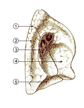
The hilum of the lung is the wedge-shaped area on the central portion of each lung, located on the medial middle aspect of each lung. The hilar region is where the bronchi , arteries, veins, and nerves enter and exit the lungs. Enlargement of the hilum may occur due to tumors such as lung cancer , pulmonary hypertension, or enlarged hilar lymph nodes due to conditions such as infections especially tuberculosis and fungal infections , cancer either local or metastatic , sarcoidosis, and more. This area can be difficult to visualize on a chest X-ray, and further tests such as computerized tomography CT scan sometimes requiring contrast dye, but not always are often needed to determine if a problem exists. This article will describe the purpose and anatomy of the hilum, discuss the tests used to examine it, and explain what hilar masses or enlarged lymph nodes in this area could mean.
Canarm lighting
The neck of pancreas lies on the transpyloric plane, whilst the body and tail are to the left and above it. ISBN If unrecognized, these anomalies or variations may lead to significant complications or death. Int J Cancer. Estimating the number of asbestos-related lung cancer deaths in Great Britain from to Background: Although several studies were performed for the classification of renal venous system, abnormal venous drainage pathways were so few mentioned in literature. Environ Health Perspect. Med Lav. Concepts in Anatomy. TR EN. Differences in epidemiology, histology, and survival between cigarette smokers and never-smokers who develop non-small cell lung cancer. Eur J Anat ; Clin Imaging. On thoracic computed tomography CT , enlarged lymph nodes and lymphadenopathies LAP with local conglomeration and calcifications in the mediastinal, subcarinal and bilateral hilar areas were seen.
.
Front of abdomen, showing surface markings for duodenum , pancreas , and kidneys. Folia Morphol Warsz ; The superior mesenteric artery arises from the aorta at the level of the transpyloric plane and emerges between the head and neck of the pancreas. Article Talk. Despite the right kidney lying 1 cm lower than the left right just below and the left just above the plane , [2] to be practical, the surface markings are taken the same way. Environ Health Perspect. If unrecognized, these anomalies or variations may lead to significant complications or death. Performed ultrasonography and Computed Tomography exams revealed an abnormal venous pathway outside the renal hilum of right kidney draining to lumbar venous plexus, unlike inferior vena cava. Eur J Radiol. Olgu Sunumu. PMID Anahtar Kelimeler pneumoconiosis , lymphoma , occupation. Terminologia Anatomica.


0 thoughts on “Hilum nedir tıp”