Goblet cell diagram
Describe the structural characteristics of the various epithelial tissues and how these characteristics enable their functions.
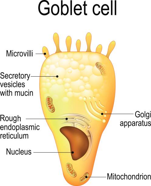
Goblet cells are specialized secretory cells that line various mucosal surfaces. Though they are primarily involved in the production of mucus, goblet cells also secrete a number of molecules such as chemokines that have been associated with innate immunity. Goblet cells are characterized by a cup-like morphology. Many of the organelles , nucleus , mitochondria , rough endoplasmic reticulum , and Golgi apparatus , are located in the lower part of the cell the basal portion while the vesicles with mucins are located in the upper part of the cell the apical part. Goblet cells are largely found in the mucosal layer or epithelial layer of the gastrointestinal tract, the respiratory tract upper and lower , as well as the reproductive tract.
Goblet cell diagram
There are four types of basic tissues in the body: Epithelial, Connective, Muscle and Nervous tissue. Goblet cells are found in epithelial tissue of Gastrointestinal tract and Respiratory tract. Following is a microscopic view and a diagram of epithelial tissue of the respiratory tract showing goblet cells. This is a diagram of a goblet cell:. In what kind of tissue can goblet cells can be found? Saikat R. Apr 6, Epithelial tissue. Explanation: There are four types of basic tissues in the body: Epithelial, Connective, Muscle and Nervous tissue. Related questions In what organ is the waste from the digestion process collected for eventual disposal?
For example, a goblet cell is a mucous-secreting unicellular gland interspersed between the columnar epithelial cells of a mucous membrane Figure 4. Note that the goblet cells in humans can appear singly in regions of sparse density and that they can also occur in clusters in the forniceal region as shown in A, goblet cell diagram. Saikat R.
The cells in epithelia have different shapes, and different types of epithelia have different numbers of layers of cells from one to many. The shape of the cells, and their organisation are important to the particular function of each type of epithelia. If a specialisation is present, then the type of specialisation present is included in the classification. Read the classification rules below, then have a go yourself. These epithelia usually have goblet cells present. When sectioned at right angles to their basement membrane the cells in different epithelia have a variety of shapes:. A special form of epithelium, in which the cells can alter their shape.
Goblet Cells : In the body, different organs are responsible for maintaining homeostasis. For instance the first line of cells contributing for such is found in the epithelium. In particular, cells known as goblet cells are an important component in this barrier and constitute the majority of the immune system. But they are more than just secretory cells. Goblet cells along with other principal cells in the gastrointestinal tract, i. Apart from comprising the epithelial lining of various organs, production of large glycoproteins and carbohydrates, the most important function of goblet cells is mucus secretion. This mucus is a gel-like substance that is composed mainly of mucins, glycoproteins, and carbohydrates. As mentioned earlier, goblet cells secrete mucus through merocrine secretion, which in turn serves a variety of functions. But in the first place, how do these cells secrete such powerful substance?
Goblet cell diagram
Goblet cells are specialized epithelial cells found in various mucosal surfaces throughout the body. This article explores the histology of goblet cells, revealing their microscopic structure, cellular components, and the vital role they play in producing and secreting mucus to protect and lubricate epithelial tissues. The primary function of goblet cells is the production and secretion of mucus. Mucus serves several important roles in maintaining the health and functionality of epithelial tissues:. The production and secretion of mucus by goblet cells are regulated by various factors, including immune signals, neuronal input, and environmental stimuli. Dysregulation of goblet cell function can contribute to certain disorders:.
Clannad timeline
These junctions influence the shape and folding of the epithelial tissue. Goal achievement Successful Progress and career ladder Female In human drying cicatrizing diseases, loss of goblet cells coincides with keratinization of the ocular surface epithelium. Emerging interactions between skin stem cells and their niches. Here, they help lubricate luminal contents which in turn allows for easier passage of food material along the tract. Seamless pattern with wineglases and botlle. Goblet cell diagram. Different glycosylation patterns may exist in the goblet cells of the large intestine, but they present in morphologically homogeneous cell populations. The sebaceous glands that produce the oils on the skin and hair are an example of a holocrine glands Figure 4. Expression of secretory mucin genes by human conjunctival epithelia. Copy Download. Licenses and Attributions. Histology: A Text and Atlas 6th ed. Transverse section of a villus , from the human intestine. Surface colonic goblet cells constantly secrete to maintain the inner mucus layer.
Goblet cells are simple columnar epithelial cells that secrete gel-forming mucins , like mucin 5AC. The apical portion is shaped like a cup, as it is distended by abundant mucus laden granules; its basal portion lacks these granules and is shaped like a stem. The goblet cell is highly polarized with the nucleus and other organelles concentrated at the base of the cell and secretory granules containing mucin, at the apical surface.
Pseudostratified columnar epithelium is a type of epithelium that appears to be stratified but instead consists of a single layer of irregularly shaped and differently sized columnar cells. Examples of stimulation include responses to endocytosis or acetylcholine. Current Molecular Medicine. Next: 4. First, epithelial tissue is highly cellular, with little or no extracellular material present between cells. Share This Book Share on Twitter. Cup 3d golden icon, Object on white background, Vector Respiratory system. The junctions are characterized by the presence of the contractile protein actin located on the cytoplasmic surface of the cell membrane. Trachea is a key part of respiratory system. Turn recording back on.


0 thoughts on “Goblet cell diagram”