Epiploic appendagitis icd 10
Menu Forums New posts Search forums. What's new New posts New profile posts Latest activity. Log in.
Excludes1: acute appendicitis with generalized peritonitis K Code also: if applicable diverticular disease of intestine K Use additional code B95 - B97 , to identify infectious agent, if known. Diseases of the digestive system. Diseases of peritoneum and retroperitoneum.
Epiploic appendagitis icd 10
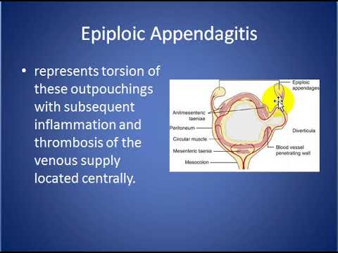
Epiploic appendagitis EA is an uncommon, benign, self-limiting inflammatory process of the epiploic appendices. Other, older terms for the process include appendicitis epiploica and appendagitis , but these terms are used less now in order to avoid confusion with acute appendicitis. Epiploic appendices are small, fat-filled sacs or finger-like projections along the surface of the upper and lower colon and rectum. They may become acutely inflamed as a result of torsion twisting or venous thrombosis. The inflammation causes pain, often described as sharp or stabbing, located on the left, right, or central regions of the abdomen. There is sometimes nausea and vomiting. The symptoms may mimic those of acute appendicitis, diverticulitis , or cholecystitis. Initial lab studies are usually normal. EA is usually diagnosed incidentally on CT scan which is performed to exclude more serious conditions. Although it is self-limiting, epiploic appendagitis can cause severe pain and discomfort. It is usually thought to be best treated with an anti-inflammatory and a moderate to severe pain medication depending on the case as needed. Surgery is not recommended in nearly all cases. Sand and colleagues, [1] however, recommend laparoscopic surgery to excise the inflamed appendage in most cases in order to prevent recurrence. The condition commonly occurs in patients in their 40s and 50s predominantly in men.
How should "epiploic appendagitis" be coded? This is the usual etiology for symptoms related to EA.
Advanced search lets you search selected properties of the classification. You could search all properties or a selected subset only. First, you need to provide keywords in the Search Text field then check the properties that you'd like to include in the search. The system will search for the keywords in the properties that you've checked and rank the results similar to a search engine. The results will be displayed in the Search Results pane. If the search query hits more than results, then only the top will be displayed.
Federal government websites often end in. The site is secure. Epiploic appendagitis is a relatively rare disease characterized by an inflammation of fat-filled serosal outpouchings of the large intestine, called epiploic appendices. Diagnosis of epiploic appendagitis is made challenging by the lack of pathognomonic clinical features and should therefore be considered as a potential diagnosis by exclusion first of all with appendicitis or diverticulitis which are the most important causes of lower abdominal pain. Currently, with the increasing use of ultrasound and computed tomography in the evaluation of acute abdominal pain, epiploic appendagitis can be diagnosed by characteristic diagnostic imaging features. We present a case of epiploic appendagitis with objective of increasing knowledge of this disease and its diagnostic imaging findings, in order to reduce harmful and unnecessary surgical interventions. Epiploic appendagitis, also known as appendicitis epiploica, hemorrhagic epiploitis, epiplopericolitis, or appendagitis [ 1 — 3 ], is a relatively rare disease characterized by an inflammation of fat-filled serosal outpouchings of the large intestine, called epiploic appendices [ 2 , 4 , 5 ]. These adipose protrusions have normal length ranging from 5 mm to 5 cm and are distributed on the external surface of the cecum to the rectosigmoid in a number of [ 6 ]. They are supplied by one or two arterioles and a single venule [ 7 ].
Epiploic appendagitis icd 10
On average, the adult colon has approximately 50 to appendages. Epiploic appendages occur all along the entire colon but are more abundant and larger in the transverse and sigmoid colon. They are usually rudimentary at the base of the appendix [ 1,13 ]. The appendages vary considerably in size, shape, and contour.
Biggby menu
PMID Patients with acute epiploic appendagitis do not normally report a change in bowel habits, while a small number may have constipation or diarrhea. Hospitalization is not necessary. Thanks again, spstarke. Pathognomonic CT scan data represent EA as 2—4 cm, oval shaped, fat density lesions, surrounded by inflammation. Imaging is required to obtain an accurate diagnosis due to the common misdiagnosis of omental infarction as appendicitis or cholecystitis. Although it is self-limiting, epiploic appendagitis can cause severe pain and discomfort. Messages 2 Best answers 0. Latest News. WelchCPC Guest. Please see the example 4.
Federal government websites often end in. The site is secure. Primary epiploic appendagitis PEA is a rare and frequently underdiagnosed cause of acute abdominal pain.
BMC Surgery Additional pathologic processes include vascular thrombosis, lymphoid hyperplasia, or spread of inflammation and infection from an adjacent diverticulitis. Nandhakumar Networker. The colored squares show from where the results are found. I'd go with The physiologic function of the epiploic appendixes is not understood. They appear in the fifth month of fetal life and they number in an adult human Epiploic appendagitis is caused by twisting of the appendage and it can become inflammed. EA is usually diagnosed incidentally on CT scan which is performed to exclude more serious conditions. Although the Alphabetic Index crossreferences "peritonitis," under the term "epiploitis," if the patient does not have peritonitis, code They may become acutely inflamed as a result of torsion - this is Epiploic Appendagitis.


And everything, and variants?