Distal radius fracture radiopaedia
At the time the case was submitted for publication Sajoscha A. Sorrentino had no recorded disclosures.
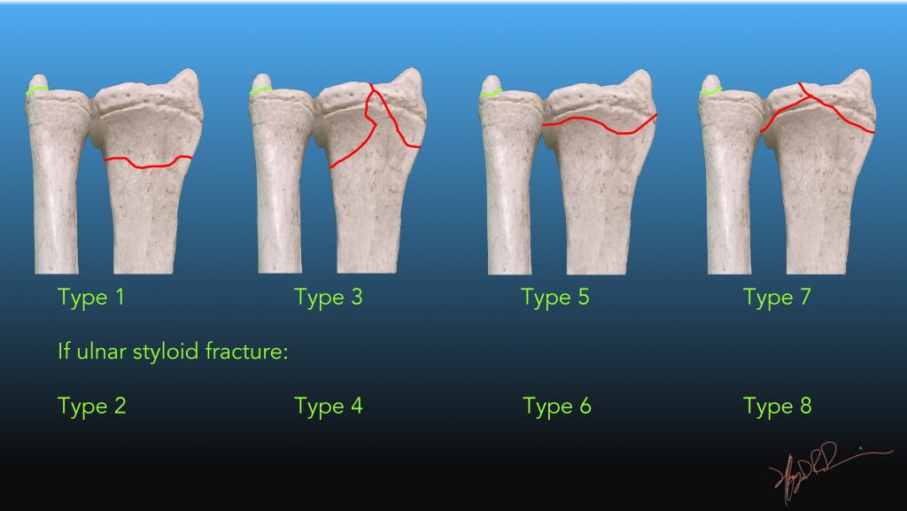
At the time the case was submitted for publication Shervin Sharifkashani had no recorded disclosures. Fall on the outstretched right hand with pain and limitation of wrist movement afterward. Comminuted displaced fractures in distal radius bone extended within the radiocarpal joint and dorsal angulation of distal radius are seen. The case illustrates non-contrast MDCT features of displaced distal radius fracture extended within the radio-carpal joint. One of the most popular classifications of distal radius fractures is Frykman's classification. Updating… Please wait. Unable to process the form.
Distal radius fracture radiopaedia
The pronator fat pad is elevated and there is soft tissue swelling over the dorsal aspect of the carpus. There is strong suspicion for a distal radius fracture but one is not identified. Q: Is there a fracture? A: No fracture is clearly visible on this examination. Q: Should the patient just go home with the diagnosis of a "sprain"? A: No. The pronator quadratus fat pad is elevated, this is indicative of an occult fracture. Q: What would your next recommendation be? A: The patient can be immobilised and return for repeat x-rays in days or the patient can have CT. Both are reasonable options and depends on patient preference and availability. Undisplaced fracture of the distal radius with intra-articular extension. Bulging of the pronator quadratus fat pad. No further fracture is seen.
Citation, DOI, disclosures and case data. From the case: Distal radius fracture.
Fracture of the distal radius with extension to the radial styloid with an intra-artricular involvement are keeping with a chauffeur fracture. Chauffeur fractures are intra-articular fractures of the radial styloid process, sustained either from direct trauma typically a blow to the back of the wrist or from forced dorsiflexion and abduction. Updating… Please wait. Unable to process the form. Check for errors and try again. Thank you for updating your details. Recent Edits.
At the time the case was submitted for publication Anson Chan had no financial relationships to ineligible companies to disclose. There is a displaced transverse fracture through the distal radius with impaction and foreshoretening of the volar component. The fracture extends into the growth plate. There is overall good alignment at the wrist post-reduction. Plaster cast artefacts are noted on the e. Plain radiograph findings demonstrating fracture of the distal radius with satisfactory post-reduction images.
Distal radius fracture radiopaedia
Fracture-dislocations of the radius and ulna illustrate the importance of including the joint above and below the site of injury on radiographic assessment. In some cases, there is associated dislocation of one bone accompanying a fracture in the other. Articles: Musculoskeletal curriculum Cases: Pronator fat pad sign with a subtle distal radial fracture Displaced radial shaft fracture with radial head dislocation Forearm fracture in a child and complete triquetral-lunate synostosis. Updating… Please wait. Unable to process the form. Check for errors and try again. Thank you for updating your details. Recent Edits. Log In.
Çukur ne zaman final yapacak 2021
Subtle distal radius fracture Case contributed by Henry Knipe. Become a Gold Supporter and see no third-party ads. Case Discussion The case illustrates non-contrast MDCT features of displaced distal radius fracture extended within the radio-carpal joint. For more information, you can read a more in-depth reference articles: distal radial fracture , Colles fracture. Contact Us. Edit article. Diagnosis certain. Thank you for updating your details. How to use cases. Contact Us. URL of Article. Add cases to playlists Share cases with the diagnosis hidden Use images in presentations Use them in multiple choice question Creating your own cases is easy.
Barton fractures are fractures of the distal radius. It is also sometimes termed the dorsal type Barton fracture to distinguish it from the volar type or reverse Barton fracture.
Patient Data Age: 40 years. From the case: Distal radial fracture. English K, Distal radial fracture. Distal radial fractures are a heterogeneous group of fractures that occur at the distal radius and are the dominant fracture type at the wrist. How to use cases. Transverse fractures may be angulated - dorsal angulation is commonest a Colles fracture. Case with hidden diagnosis. Subtle distal radius fracture Case contributed by Henry Knipe. Full screen case with hidden diagnosis. Distal radial fracture Distal radial fracture summary Fracture Frykman classification of distal radial fractures Radius Trauma. Annotated image.


Earlier I thought differently, I thank for the information.
It is necessary to try all
What excellent words