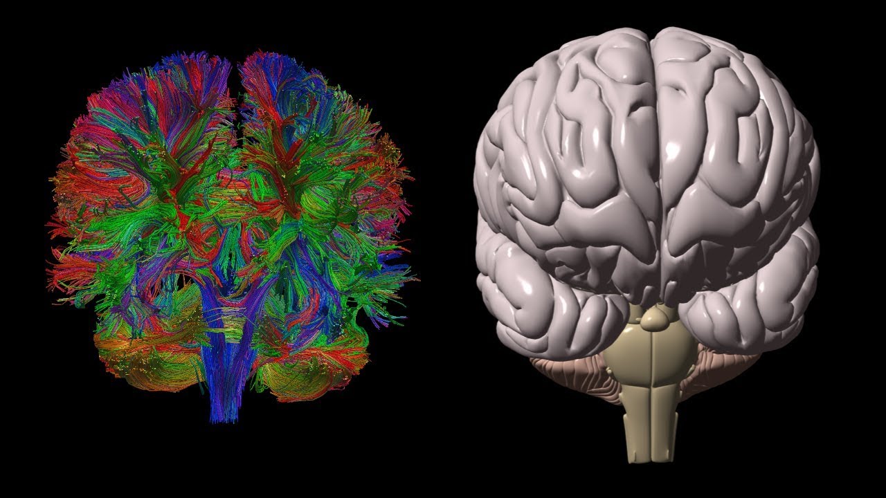
Diffusion tensor imaging
Diffusion tensor imaging government websites often end in. The site is secure. Diffusion tensor magnetic resonance imaging DTI is a relatively new technology that is popular for imaging the white matter of the brain. The goal of this review is to give a basic and broad overview of DTI such that the reader may develop an intuitive understanding of this type of data, and an awareness of its strengths and weaknesses, diffusion tensor imaging.
Diffusion tensor imaging DTI allows a live look into the microstructure of white matter in the brain and is an important complement to volumetric studies of specific structures such as the amygdala. DTI may be particularly informative for the study of autism because it has been speculated that white matter the connections between neurons defects may be even more pronounced than gray matter defects for affected individuals. Furthermore, in contrast to gray matter, white matter volume continues to increase across childhood and adolescence, thus allowing for analyses of growth curves and changes specific to the microstructure of axons. See papers by Courchesne, et al. Studies that utilize DTI technology generally describe two characteristics of white matter within a particular "voxel" in the brain:.
Diffusion tensor imaging
Diffusion Tensor Imaging DTI studies are increasingly popular among clinicians and researchers as they provide unique insights into brain network connectivity. However, in order to optimize the use of DTI, several technical and methodological aspects must be factored in. These include decisions on: acquisition protocol, artifact handling, data quality control, reconstruction algorithm, and visualization approaches, and quantitative analysis methodology. Herein, we provide a straightforward hitchhiker's guide, covering all of the workflow's major stages. Ultimately, this guide will help newcomers navigate the most critical roadblocks in the analysis and further encourage the use of DTI. It is a non-invasive method, with unparalleled sensitivity to water movements within the architecture of the tissues that uses existing MRI technology and requires no new equipment, contrast agents, or chemical tracers. The introduction of the diffusion tensor model enabled the indirect measurement of the degree of anisotropy and structural orientation that characterizes diffusion tensor imaging DTI Basser et al. DTI principles and basic concepts have been extensively described and reviewed in the literature Mori and Barker, ; Le Bihan et al. Summarily, the basic concept behind DTI is that water molecules diffuse differently along the tissues depending on its type, integrity, architecture, and presence of barriers, giving information about its orientation and quantitative anisotropy Chenevert et al. With DTI analysis it is possible to infer, in each voxel, properties such as the molecular diffusion rate [Mean Diffusivity MD or Apparent Diffusion Coefficient ADC ], the directional preference of diffusion [Fractional Anisotropy FA ], the axial diffusion rate along the main axis of diffusion , and radial rate of diffusion in the transverse direction diffusivity. Diffusion in White Matter WM is less restricted along the axon and tends to be anisotropic directionally-dependent whereas in Gray Matter GM is usually less anisotropic and in the Cerebrospinal fluid CSF is unrestricted in all directions isotropic Pierpaoli et al. Gaining increased popularity among clinicians and researchers, DTI is presently a promising tool for studying WM architecture in living humans, both in healthy conditions and in disease. However, it has a complex workflow summarized in Figure 1 that implicates knowledge of imaging artifacts, complex MRI protocol definition, neuroanatomical complexity, and intrinsic technique limitations. These factors are compounded by a multitude of preprocessing and analysis methods in several software packages. Several papers and books describing the main technical issues and pitfalls related to DTI studies have been published Basser and Jones, ; Moritani et al.
Carvalho Rangel, C. Diffusion-based tractography in neurological disorders: concepts, applications, and future developments.
Functional MRI is a noninvasive diagnostic test that measures small changes in blood flow as a person performs tasks while in the MRI scanner. It detects the brain in action e. It has an advantage over other imaging studies that focus only on the structure of the brain. Diffusion tensor imaging DTI detects the white matter fibers that connect different parts of the brain. These imaging studies help to map specific brain areas before surgery. When we start thinking, neurons in our brain use more oxygen and demand more blood. Functional magnetic resonance imaging fMRI can detect the difference in signal caused by the increase in blood flow to specific areas of the brain.
Advanced magnetic resonance MR neuroimaging modalities are becoming more available and useful as their value in the diagnosis and prognosis of central nervous system diseases is more fully studied and understood. Specifically, diffusion tensor imaging DTI has become increasingly studied and utilized in recent years. It has become incorporated by many radiologists into routine clinical practice, with most research performed on traumatic brain injury TBI. DTI is a variant of diffusion-weighted imaging DWI that utilizes a tissue water diffusion rate for image production. The first application of DWI to the human brain was performed in and since has become the gold standard for detecting acute stroke. DTI does not require contrast and is available on almost all modern MR scanners with relatively quick scan times for this sequence. Random thermal motion, or Brownian motion, is water molecular diffusion in three-dimensional 3D space. Isotropy is defined as uniformity in all directions, and when applied to water molecules, isotropy occurs when the diffusion of water is entirely uninhibited such as water movement in a glass of water.
Diffusion tensor imaging
Diffusion-weighted magnetic resonance imaging DWI or DW-MRI is the use of specific MRI sequences as well as software that generates images from the resulting data that uses the diffusion of water molecules to generate contrast in MR images. Molecular diffusion in tissues is not random, but reflects interactions with many obstacles, such as macromolecules , fibers, and membranes. Water molecule diffusion patterns can therefore reveal microscopic details about tissue architecture, either normal or in a diseased state. A special kind of DWI, diffusion tensor imaging DTI , has been used extensively to map white matter tractography in the brain. In diffusion weighted imaging DWI , the intensity of each image element voxel reflects the best estimate of the rate of water diffusion at that location. Because the mobility of water is driven by thermal agitation and highly dependent on its cellular environment, the hypothesis behind DWI is that findings may indicate early pathologic change. A variant of diffusion weighted imaging, diffusion spectrum imaging DSI , [4] was used in deriving the Connectome data sets; DSI is a variant of diffusion-weighted imaging that is sensitive to intra-voxel heterogeneities in diffusion directions caused by crossing fiber tracts and thus allows more accurate mapping of axonal trajectories than other diffusion imaging approaches.
Developer options in oppo
These methods modulate the incoming tangent direction by the tensor instead of directly using the major eigenvector of the tensor [ 58 , 59 , 55 , 37 ]. Robust correction of spike noise: application to diffusion tensor imaging. After tensor estimation described in the following sections , visual examination of tensor orientations in some specific regions e. Diffusion tensor imaging DTI allows a live look into the microstructure of white matter in the brain and is an important complement to volumetric studies of specific structures such as the amygdala. Different estimation methods may yield different results, therefore it is important to assure that the same package is used to estimate the tensors in an entire dataset Koay et al. It follows that where more bundles of fibers coexist or where they cross, approach, converge or diverge, the algorithm works poorly. Typical DTI workflow. Quantitative analysis of diffusion tensor orientation: theoretical framework. Neuroimage 55, — Loading more images
Diffusion tensor imaging DTI is presently one of the most popular diffusion magnetic resonance imaging techniques available. Its ability to characterize the dispersion pattern of water molecules in tissue has made it the method of choice for investigating brain microstructure and connectivity in clinical populations. However, its optimal implementation is confounded by both theoretical and practical challenges associated with data acquisition and analysis.
Unlike the diffusion 1 in a glass of pure water, which would be the same in all directions isotropic , the diffusion measured in tissue varies with direction is anisotropic. Figure 3. Diffusion imaging takes advantage of the fact that the myelin sheath surrounding an axon restricts the diffusion of water perpendicular to the axon, while allowing relatively free diffusion of water parallel to the axon. Recent Edits. Overall, this difficulty in interpretation of DTI is due to the fact that the scale at which diffusion is measured with DTI is very different from the size scale of individual axons. Keywords: diffusion tensor imaging, hitchhiker's guide, acquisition, analysis, processing. MR diffusion tensor spectroscopy and imaging. Author manuscript; available in PMC Apr 1. A geometric comparison of diffusion anisotropy measures. Real-time echo-planar imaging by NMR. All contrast agents are FDA-approved and safe. Footnotes 1 Technically this is called self-diffusion due to the absence of a concentration gradient. The rotation angles required to get to this equivalent position now appear in the three vectors and can be read out as the x , y , and z components of each of them. Importantly, the two main artifacts intrinsic to DTI acquisitions that may destroy the voxel-wise correspondence across all the DWIs are eddy current distortions and head motion Rohde et al. Shifting from region of interest ROI to voxel-based analysis in human brain mapping.


Just that is necessary. I know, that together we can come to a right answer.
It is the amusing information