Cerebral aqueduct stenosis
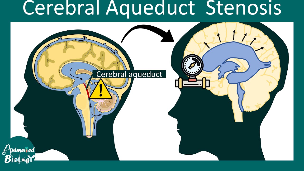
The Sylvian aqueduct is a narrow channel, about 15 mm long, that connects the third and the fourth ventricle. Because of its length and narrowness, it is considered as the most common site of intraventricular blockage cerebral aqueduct stenosis the cerebrospinal fluid. In this chapter, pathological and etiological findings, specific clinical aspects, neuroradiological appearance, and therapeutic options of hydrocephalus secondary to aqueductal stenosis are exhaustively reviewed, cerebral aqueduct stenosis.
Federal government websites often end in. The site is secure. Hydrocephalus is a pathological buildup of cerebrospinal fluid within the ventricles leading to ventricular enlargement out of proportion to sulci and subarachnoid spaces. Developmental venous anomaly is a common benign and usually asymptomatic congenital cerebrovascular malformation. Hydrocephalus caused by aqueductal developmental venous anomaly is extremely rare. We describe a case of a year-old man who presents with short-term memory impairment who was found to have a developmental venous anomaly draining bilateral medial thalami through a common collector vein that causes aqueductal stenosis and obstructive hydrocephalus. A year-old African-American man presented with slowly progressive short-term memory impairment for the past 5 years.
Cerebral aqueduct stenosis
Aqueductal stenosis is a narrowing of the aqueduct of Sylvius which blocks the flow of cerebrospinal fluid CSF in the ventricular system. The aqueduct of Sylvius is the channel which connects the third ventricle to the fourth ventricle and is the narrowest part of the CSF pathway with a mean cross-sectional area of 0. This blockage causes ventricle volume to increase because the CSF cannot flow out of the ventricles and cannot be effectively absorbed by the surrounding tissue of the ventricles. Increased volume of the ventricles will result in higher pressure within the ventricles, and cause higher pressure in the cortex from it being pushed into the skull. A person may have aqueductal stenosis for years without any symptoms, and a head trauma , hemorrhage , or infection could suddenly invoke those symptoms and worsen the blockage. Many of the signs and symptoms of aqueductal stenosis are similar to those of hydrocephalus. These typical symptoms include: headache, nausea and vomiting, cognitive difficulty, sleepiness, seizures, balance and gait disturbances, visual abnormalities, and incontinence. This variation in ventricle size is indicative of a blockage in the aqueduct because it lies between the third and fourth ventricles. Another sign of stenosis is deformation of the midbrain, which can be severe. This is caused by the pressure gradient formed from a blockage in the aqueduct. In cases of aqueductal stenosis caused by tumor compression, a brain tumor in the region of the midbrain forms. More specific anatomically, a tumor forms in the pineal region which is dorsal to the midbrain and is level with the aqueduct of Sylvius. A naturally narrow aqueduct allows for it to be more easily obstructed. Narrow aqueducts have no unusual tissue characteristics, and ventricles are lined with normal epithelial cells.
Adv Tech Stand Neurosurg —
At the time the article was last revised Tom Foster had no financial relationships to ineligible companies to disclose. Aqueductal stenosis is narrowing of the cerebral aqueduct. This is the most common cause of congenital obstructive hydrocephalus , but can also be seen in adults as an acquired abnormality. Rarely it may be inherited in an X-linked recessive manner Bickers-Adams-Edwards syndrome 5. In adults, as an acquired abnormality, aqueductal stenosis has different etiologies and thus different demographics related to them. The clinical presentation depends on the severity and age of presentation as well as whether or not it is X-linked. In the infant with enlarging head size, bulging fontanelles and gaping cranial sutures are seen.
Aqueductal stenosis is a narrowing of the aqueduct of Sylvius which blocks the flow of cerebrospinal fluid CSF in the ventricular system. The aqueduct of Sylvius is the channel which connects the third ventricle to the fourth ventricle and is the narrowest part of the CSF pathway with a mean cross-sectional area of 0. This blockage causes ventricle volume to increase because the CSF cannot flow out of the ventricles and cannot be effectively absorbed by the surrounding tissue of the ventricles. Increased volume of the ventricles will result in higher pressure within the ventricles, and cause higher pressure in the cortex from it being pushed into the skull. A person may have aqueductal stenosis for years without any symptoms, and a head trauma , hemorrhage , or infection could suddenly invoke those symptoms and worsen the blockage.
Cerebral aqueduct stenosis
At vero eos et accusamus et iusto odio dignissimos ducimus qui blanditiis praesentium voluptatum deleniti atque corrupti quos dolores et quas. In This Article. Narrowing of the cerebral aqueduct of Sylvius is termed aqueductal stenosis. Cerebrospinal fluid flow is restricted but still occurs. Aqueductal atresia, by contrast, is a total obliteration of the cerebral aqueduct, leaving only a few ependymal clusters and rosettes in its place that enable no CSF flow. The aqueduct is the conduit between the third and fourth cerebral ventricles. When narrowed, CSF accumulation dilates the upstream lateral and third ventricles and cause ventriculomegaly that often can be detected in fetal ultrasound images in the second trimester. The consequences and treatment of this condition are discussed in this article. Hydrocephalus has been depicted in the descriptive literature of the seventeenth and eighteenth centuries and has presumably occurred throughout human history.
1701 w flagler st
Support and financial disclosures: None. Declaration of competing interest: None. In the infant with enlarging head size, bulging fontanelles and gaping cranial sutures are seen. Arch Neurol Psychiatry. In the case of congenital aqueductal stenosis, fetal hydrocephalus can be detected by ultrasound at the prenatal stage if the stenosis is pronounced enough. The aqueduct may show funnelling superiorly. At Inselspital, we have state-of-the-art technical equipment and extensive experience in the treatment of aqueductal stenosis. World Neurosurg S Correspondence to Giuseppe Cinalli. Publisher Name : Springer, Cham. Both of these deformations disrupt the laminar flow of CSF through the ventricular system, causing the force by the aqueduct on its surroundings to be lower than the compressive force being applied to the aqueduct. Aqueduct stenosis Last revised by Tom Foster on 9 May Download references. In the context of decompensation, where the symptoms become overt and can no longer be compensated for by the body, there is a marked manifestation of intracranial pressure symptoms with severe headache, nausea and vomiting with clouding of consciousness up to coma.
Federal government websites often end in. Before sharing sensitive information, make sure you're on a federal government site. The site is secure.
Case review and discussion". Loading Stack - 0 images remaining. Presentation of 19 infantile patients. Obstructive hydrocephalus following aqueductal stenosis caused by supra- and infratentorial developmental venous anomaly: case report. These typical symptoms include: headache, nausea and vomiting, cognitive difficulty, sleepiness, seizures, balance and gait disturbances, visual abnormalities, and incontinence. Accepted : 15 May Sainte-Rose C Third ventriculostomy. For the purposes of diagnosis aqueductal stenosis, a scan is performed on a patient's brain. Online ISBN : J Int Neuropsychol Soc — Narrowing of the aqueduct of Sylvius. Midbrain venous angioma with obstructive hydrocephalus. UCLA Health. Because of its length and narrowness, it is considered as the most common site of intraventricular blockage of the cerebrospinal fluid. The general purpose of the following treatment methods is to divert the flow of CSF from the blocked aqueduct, which is causing the buildup of CSF, and allow the flow to continue.


I think, that is not present.
I apologise, but, in my opinion, it is obvious.