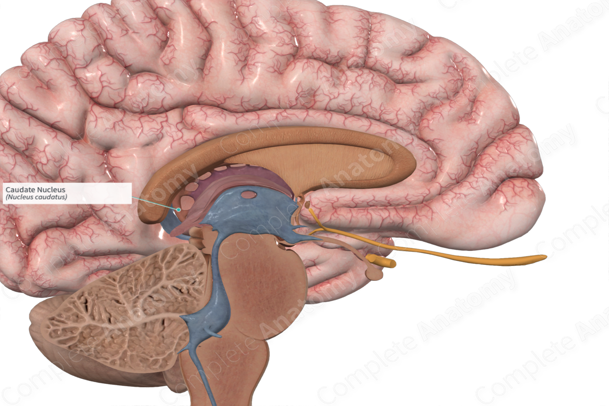
Caudate nucleus
Deep within each half of the brain lies the caudate nucleus.
Federal government websites often end in. Before sharing sensitive information, make sure you're on a federal government site. The site is secure. NCBI Bookshelf. Margaret E. Driscoll ; Pradeep C.
Caudate nucleus
At the time the article was last revised Yoshi Yu had no financial relationships to ineligible companies to disclose. Caudate nuclei are paired nuclei which along with the globus pallidus and putamen are referred to as the corpus striatum , and collectively make up the basal ganglia. The caudate nuclei have both motor and behavioral functions, in particular maintaining body and limb posture, as well as controlling approach-attachment behaviors, respectively 3. The caudate nucleus is located lateral to the lateral ventricles, with the head lateral to the frontal horn, and body lateral to the body of the lateral ventricle. The tail of the caudate nucleus terminates immediately above the temporal horn of the ventricle. It is bound laterally by the anterior crus of the internal capsule. The head of the caudate nucleus is supplied by the recurrent artery of Heubner , a small branch from the A2 sometimes the A1 segment of the anterior cerebral artery. The superior aspect of the head and the body of the caudate are supplied by the lenticulostriate perforators from the middle cerebral artery. The tail of the caudate is supplied by the anterior choroidal artery. Please Note: You can also scroll through stacks with your mouse wheel or the keyboard arrow keys. Updating… Please wait. Unable to process the form. Check for errors and try again. Thank you for updating your details.
Caudate nucleus stroke presents with nonprogressive symptoms, caudate nucleus, which stabilize in severity less than one hour after onset. Some highlights of research on the effects of caudate nucleus lesions over the past years.
It plays a critical role in various higher neurological functions. Each caudate nucleus is composed of a large anterior head, a body, and a thin tail that wraps anteriorly such that the caudate nucleus head and tail can be visible in the same coronal cut. When combined with the putamen, the pair is referred to as the striatum and is often considered jointly in function. The striatum is the major input source for the basal ganglia, which also includes the globus pallidus, subthalamic nucleus, and substantia nigra. These deep brain structures together largely control voluntary skeletal movement.
The caudate nucleus is one of the structures that make up the corpus striatum , which is a component of the basal ganglia in the human brain. The caudate is also one of the brain structures which compose the reward system and functions as part of the cortico — basal ganglia — thalamic loop. Together with the putamen , the caudate forms the dorsal striatum , which is considered a single functional structure; anatomically, it is separated by a large white matter tract, the internal capsule , so it is sometimes also referred to as two structures: the medial dorsal striatum the caudate and the lateral dorsal striatum the putamen. In this vein, the two are functionally distinct not as a result of structural differences, but merely due to the topographical distribution of function. The caudate nuclei are located near the center of the brain, sitting astride the thalamus. There is a caudate nucleus within each hemisphere of the brain. Individually, they resemble a C-shape structure with a wider "head" caput in Latin at the front, tapering to a "body" corpus and a "tail" cauda. Sometimes a part of the caudate nucleus is referred to as the "knee" genu. The head and body of the caudate nucleus form part of the floor of the anterior horn of the lateral ventricle.
Caudate nucleus
Deep within each half of the brain lies the caudate nucleus. The caudate nucleus is a pair of brain structures that make up part of the basal ganglia. It helps control high-level functioning, including:.
Cactus jacks percentage
The anterior portion of the caudate nucleus is connected with the lateral and medial prefrontal cortices and is involved in working memory and executive functioning. Specifically, a larger caudate nucleus volume has been linked with better verbal fluency performance. The caudate may contribute to a variety of other cognitive functions as well, ranging from habit learning to attention. The ventral telencephalon gives rise to the lateral and medial ganglionic eminences, which are the source of neurons in the striatum and cortical interneurons, respectively. Caudate nucleus volumes and genetic determinants of homocysteine metabolism in the prediction of psychomotor speed in older persons with depression. Case report. The Journal of Neuroscience. Sexual dimorphism of brain developmental trajectories during childhood and adolescence. Clinical diagnosis of subcortical infarction, chiefly lacunar stroke, [ Search term. View a 3-D diagram and learn about related…. Haber S. Bilateral lesions in the head of the caudate nucleus in cats were correlated with a decrease in the duration of deep slow wave sleep during the sleep-wakefulness cycle. The caudate is also one of the brain structures which compose the reward system and functions as part of the cortico — basal ganglia — thalamic loop.
Watching a movie can be a mesmerizing experience, not just for our eyes — but also for our ears. The most powerful scene in Maestro is arguably one in which there is no dialogue — only music. Trumpets pierce the air.
Expert Rev Neurother. Article Talk. Caudate nucleus volumes in frontotemporal lobar degeneration: differential atrophy in subtypes. Figure 8a: coronal Gray's illustrations Figure 8a: coronal Gray's illustrations. Figure 2: brainstem nuclei and their connections Figure 2: brainstem nuclei and their connections. We experience these things every day, but how do our brains create them? However, in the CNS, a glymphatic system removes waste from the CSF and allows it to drain to the periphery rather than a lymphatic system. For this reason, lacunar infarction should be regarded as a potentially severe condition rather than a relatively benign disorder and, therefore, lacunar stroke patients require adequate and rigorous management and follow-up. Structure of the striatum in the basal ganglia of the brain. Log in Sign up. The tail of the caudate nucleus terminates immediately above the temporal horn of the ventricle. Hum Brain Mapp. Annual Review of Neuroscience. Know Your Brain: Striatum.


I here am casual, but was specially registered to participate in discussion.
Rather curious topic
You are not right. Write to me in PM, we will talk.