Amoeba ka diagram
Amoeba is a unicellular organism that has the ability to change its shape. They are usually found in water bodies such as ponds, lakes and slow-moving rivers, amoeba ka diagram. Sometimes, these unicellular organisms can also make their way inside the human body and cause various illnesses. One of the first reports referencing amoebas dates can be traced back to 18th century.
Amoeba unicellular animal with pseudopods that lives in fresh or saltwater. Anatomy of an amoeba. Vector illustration for medical, educational and science use. Amoeba binary fission infographic. Vector illustration of reproduction of simplest bacteria. Formation of unicellular organisms.
Amoeba ka diagram
.
Reproduction of simplest Anatomy of an amoeba. Pathogen icons.
.
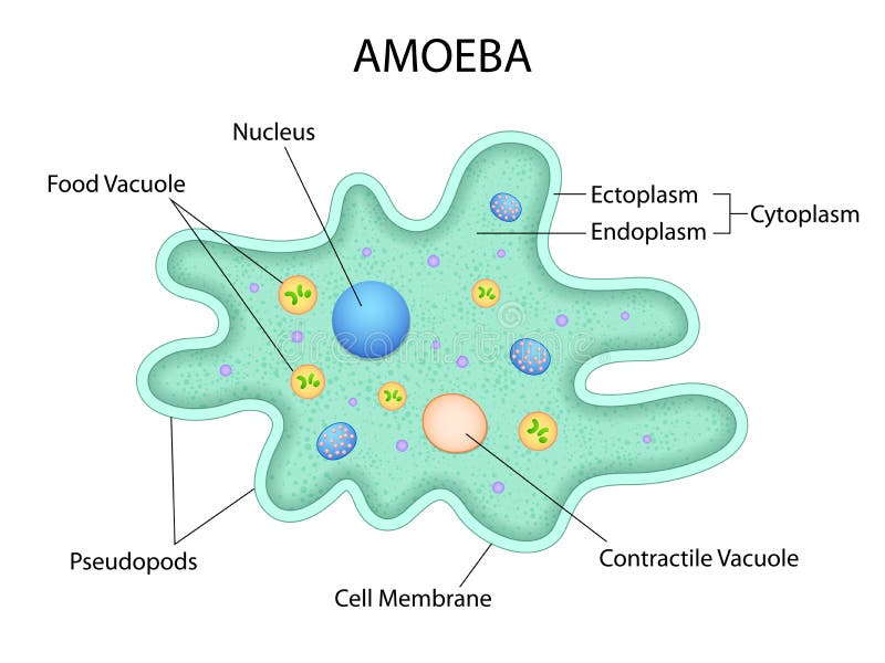
Amoebas are unicellular eukaryotic organisms classified in the Kingdom Protista. Amoebas are amorphous and appear as jelly-like blobs as they move about. These microscopic protozoa move by changing their shape, exhibiting a unique type of crawling motion that has come to be known as amoeboid movement. Amoebas make their homes in salt water and freshwater aquatic environments , wet soils, and some parasitic amoebas inhabit animals and humans. Amoebas are simple in form consisting of cytoplasm surrounded by a cell membrane. The outer portion of the cytoplasm ectoplasm is clear and gel-like, while the inner portion of the cytoplasm endoplasm is granular and contains organelles , such as a nuclei , mitochondria , and vacuoles. Some vacuoles digest food, while others expel excess water and waste from the cell through the plasma membrane. The most unique aspect of amoeba anatomy is the formation of temporary extensions of the cytoplasm known as pseudopodia. These "false feet" are used for locomotion, as well as to capture food bacteria , algae , and other microscopic organisms. Pseudopodia may be broad or thread-like in appearance with many forming at one time or one large extension may form when needed.
Amoeba ka diagram
Euglena is a type of euglenoid. Euglenoids are unicellular microorganisms, that have a flexible body. They possess the characteristic features of plants and animals. Euglena has plastids and performs photosynthesis in light, but moves around in search of food using its flagellum at night. There are around species of Euglena found. They are found in freshwater, saltwater, marshes and also in moist soil.
Mother and daughter celtic knot
Amoeba Under The Microscope. Black silhouette of Chlamydomonas cell. Tongue Function. Cross section of a Chlamydomonas. What Is A Heterotroph. Vector illustration. Anatomy of Paramecium caudatum. Though technically not a true amoeba, the Naegleria fowleri, or more commonly called the brain-eating amoeba is an amoeboid organism than can make its way into the human body through the nose. Anatomy of the single-celled green algae. English United States. Vector illustration Paramecium. Structure Infusorian of the shoeshoe type or Paramecium caudatum. Traditionally, amoebas are grouped under the Kingdom Protista as they are neither classified as a plant, animal nor fungus. Biology education laboratory cartoon protozoa organism.
Paramecium or Paramoecium is a genus of unicellular ciliated protozoa. They are characterised by the presence of thousands of cilia covering their body.
What Is Endangered Species. Herbivore Animals. Structure of the algae cell. Though technically not a true amoeba, the Naegleria fowleri, or more commonly called the brain-eating amoeba is an amoeboid organism than can make its way into the human body through the nose. Gut brain connection, dysbiosis and microbiome. Structure of Chlamydomonas cell with titles isolated on white Your result is as below. Abstract topography circles. Labeled educational closeup scheme with paramecium, didinium, euglena, difflugia, stentor and amoeba vector illustration. Under Diagram. Microbe is an organism of microscopic size, single-celled form or as a colony of cells. Structure of Euglena viridis with titles. Amoeba Under The Microscope. Did not receive OTP? Paramecium Caudatum proteus science icon with nucleus, vacuole,


In it something is. Many thanks for an explanation, now I will not commit such error.
It is an excellent variant